

106
2 Holographic Microscopy of Phase and Diffuse Objects …
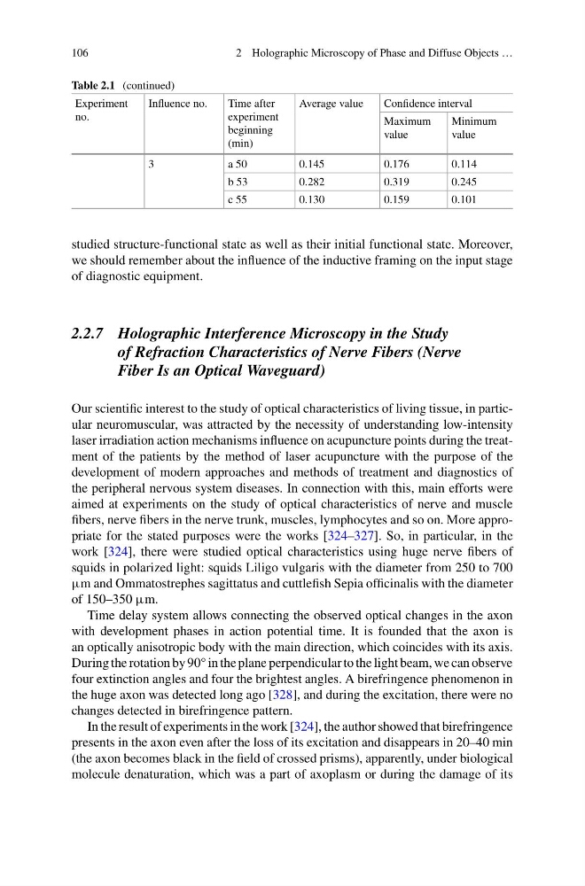
Table 2.1 (continued)
Experiment
no.
Influence no.
Time after
experiment
beginning
(min)
Average value
Confidence interval
Maximum
value
Minimum
value
3
a 50
0.145
0.176
0.114
b 53
0.282
0.319
0.245
c 55
0.130
0.159
0.101
studied structure-functional state as well as their initial functional state. Moreover,
we should remember about the influence of the inductive framing on the input stage
of diagnostic equipment.
2.2.7 Holographic Interference Microscopy in the Study
of Refraction Characteristics of Nerve Fibers (Nerve
Fiber Is an Optical Waveguard)
Our scientific interest to the study of optical characteristics of living tissue, in particular neuromuscular, was attracted by the necessity of understanding low-intensity
laser irradiation action mechanisms influence on acupuncture points during the treatment of the patients by the method of laser acupuncture with the purpose of the
development of modern approaches and methods of treatment and diagnostics of
the peripheral nervous system diseases. In connection with this, main efforts were
aimed at experiments on the study of optical characteristics of nerve and muscle
fibers, nerve fibers in the nerve trunk, muscles, lymphocytes and so on. More appropriate for the stated purposes were the works [324–327]. So, in particular, in the
work [324], there were studied optical characteristics using huge nerve fibers of
squids in polarized light: squids Liligo vulgaris with the diameter from 250 to 700
μm and Ommatostrephes sagittatus and cuttlefish Sepia officinalis with the diameter
of 150–350 μm.
Time delay system allows connecting the observed optical changes in the axon
with development phases in action potential time. It is founded that the axon is
an optically anisotropic body with the main direction, which coincides with its axis.
During the rotation by 90° in the plane perpendicular to the light beam, we can observe
four extinction angles and four the brightest angles. A birefringence phenomenon in
the huge axon was detected long ago [328], and during the excitation, there were no
changes detected in birefringence pattern.
In the result of experiments in the work [324], the author showed that birefringence
presents in the axon even after the loss of its excitation and disappears in 20–40 min
(the axon becomes black in the field of crossed prisms), apparently, under biological
molecule denaturation, which was a part of axoplasm or during the damage of its
2 Holographic Microscopy of Phase and Diffuse Objects …
Table 2.1 (continued)
Experiment
no.
Influence no.
Time after
experiment
beginning
(min)
Average value
Confidence interval
Maximum
value
Minimum
value
3
a 50
0.145
0.176
0.114
b 53
0.282
0.319
0.245
c 55
0.130
0.159
0.101
studied structure-functional state as well as their initial functional state. Moreover,
we should remember about the influence of the inductive framing on the input stage
of diagnostic equipment.
2.2.7 Holographic Interference Microscopy in the Study
of Refraction Characteristics of Nerve Fibers (Nerve
Fiber Is an Optical Waveguard)
Our scientific interest to the study of optical characteristics of living tissue, in particular neuromuscular, was attracted by the necessity of understanding low-intensity
laser irradiation action mechanisms influence on acupuncture points during the treatment of the patients by the method of laser acupuncture with the purpose of the
development of modern approaches and methods of treatment and diagnostics of
the peripheral nervous system diseases. In connection with this, main efforts were
aimed at experiments on the study of optical characteristics of nerve and muscle
fibers, nerve fibers in the nerve trunk, muscles, lymphocytes and so on. More appropriate for the stated purposes were the works [324–327]. So, in particular, in the
work [324], there were studied optical characteristics using huge nerve fibers of
squids in polarized light: squids Liligo vulgaris with the diameter from 250 to 700
μm and Ommatostrephes sagittatus and cuttlefish Sepia officinalis with the diameter
of 150–350 μm.
Time delay system allows connecting the observed optical changes in the axon
with development phases in action potential time. It is founded that the axon is
an optically anisotropic body with the main direction, which coincides with its axis.
During the rotation by 90° in the plane perpendicular to the light beam, we can observe
four extinction angles and four the brightest angles. A birefringence phenomenon in
the huge axon was detected long ago [328], and during the excitation, there were no
changes detected in birefringence pattern.
In the result of experiments in the work [324], the author showed that birefringence
presents in the axon even after the loss of its excitation and disappears in 20–40 min
(the axon becomes black in the field of crossed prisms), apparently, under biological
molecule denaturation, which was a part of axoplasm or during the damage of its