

6 Bio-microelectromechanical Systems (BioMEMS) …
143
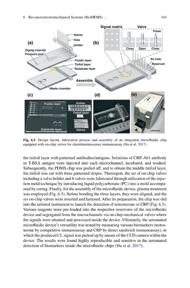
Fig. 6.5 Design layout, fabrication process and assembly of an integrated microfluidic chip
equipped with on-chip valves for chemiluminescence immunoassay (Hu et al. 2017)
the tinfoil layer with patterned antibodies/antigens. Solutions of CRP-Ab1 antibody
or T-BSA antigen were injected into each microchannel, incubated, and washed.
Subsequently, the PDMS chip was peeled off, and to obtain the middle tinfoil layer,
the tinfoil was cut with three patterned stripes. Thereupon, the set of on-chip valves
including a valve holder and 6 valves were fabricated through utilization of the injection mold technique by introducing liquid polycarbonate (PC) into a mold accompanied by curing. Finally, for the assembly of the microfluidic device, plasma treatment
was employed (Fig. 6.5). Before bonding the three layers, they were aligned, and the
six on-chip valves were inserted and fastened. After its preparation, the chip was slid
into the tailored instrument to launch the detection of testosterone or CRP (Fig. 6.5).
Various reagents were pre-loaded into the respective reservoirs of the microfluidic
device and segregated from the microchannels via on-chip mechanical valves where
the signals were attained and processed inside the device. Ultimately, the automated
microfluidic device’s versatility was tested by measuring various biomarkers (testosterone by competitive immunoassay and CRP by direct sandwich immunoassay), in
which the produced CL signal was picked up by means of the CCD camera within the
device. The results were found highly reproducible and sensitive in the automated
detection of biomarkers inside the microfluidic chips (Hu et al. 2017).
143
Fig. 6.5 Design layout, fabrication process and assembly of an integrated microfluidic chip
equipped with on-chip valves for chemiluminescence immunoassay (Hu et al. 2017)
the tinfoil layer with patterned antibodies/antigens. Solutions of CRP-Ab1 antibody
or T-BSA antigen were injected into each microchannel, incubated, and washed.
Subsequently, the PDMS chip was peeled off, and to obtain the middle tinfoil layer,
the tinfoil was cut with three patterned stripes. Thereupon, the set of on-chip valves
including a valve holder and 6 valves were fabricated through utilization of the injection mold technique by introducing liquid polycarbonate (PC) into a mold accompanied by curing. Finally, for the assembly of the microfluidic device, plasma treatment
was employed (Fig. 6.5). Before bonding the three layers, they were aligned, and the
six on-chip valves were inserted and fastened. After its preparation, the chip was slid
into the tailored instrument to launch the detection of testosterone or CRP (Fig. 6.5).
Various reagents were pre-loaded into the respective reservoirs of the microfluidic
device and segregated from the microchannels via on-chip mechanical valves where
the signals were attained and processed inside the device. Ultimately, the automated
microfluidic device’s versatility was tested by measuring various biomarkers (testosterone by competitive immunoassay and CRP by direct sandwich immunoassay), in
which the produced CL signal was picked up by means of the CCD camera within the
device. The results were found highly reproducible and sensitive in the automated
detection of biomarkers inside the microfluidic chips (Hu et al. 2017).