

176
8 Morphogenesis
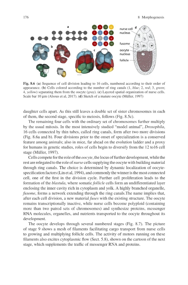
Fig. 8.6 (a) Sequence of cell division leading to 16 cells, numbered according to their order of
appearance. (b) Cells colored according to the number of ring canals (1, blue; 2, red; 3, green;
4, yellow) separating them from the oocyte (gray). (c) Layered spatial organization of nurse cells.
Scale bar 10 μm (Alsous et al, 2017). (d) Sketch of a mature oocyte (Müller, 1997)
daughter cells apart. As this still leaves a double set of sister chromosomes in each
of them, the second stage, specific to meiosis, follows (Fig. 8.5c).
The remaining four cells with the ordinary set of chromosomes further multiply
by the usual mitosis. In the most intensively studied “model animal”, Drosophila,
16 cells connected by thin tubes, called ring canals, form after two more divisions
(Fig. 8.6a and b). Four divisions prior to the onset of specialization is a conserved
feature among animals; also in mice, far ahead on the evolution ladder and a proxy
for humans in genetic studies, roles of cells begin to diversify from the 12 to16 cell
stage (Müller, 1997).
Cells compete for the role of the oocyte, the locus of further development, while the
rest are relegated to the role of nurse cells supplying the oocyte with building material
through ring canals. The choice is determined by dynamic localization of oocytespecification factors (Lin et al, 1994), and commonly the winner is the most connected
cell, one of the first in the division cycle. Further cell proliferation leads to the
formation of the blastula, where somatic follicle cells form an undifferentiated layer
enclosing the inner cavity rich in cytoplasm and yolk. A highly branched organelle,
fusome, forms a network extending through the ring canals.The name implies that,
after each cell division, a new material fuses with the existing structure. The oocyte
remains transcriptionally inactive, while nurse cells become polyploid (containing
more than two paired sets of chromosomes) and synthesize proteins, messenger
RNA molecules, organelles, and nutrients transported to the oocyte throughout its
development.
The oocyte develops through several numbered stages (Fig. 8.7). The picture
of stage 9 shows a mesh of filaments facilitating cargo transport from nurse cells
to growing and multiplying follicle cells. The activity of motors running on these
filaments also excites cytoplasmic flow (Sect. 5.8), shown on the cartoon of the next
stage, which supplements the traffic of messenger RNA and proteins.
8 Morphogenesis
Fig. 8.6 (a) Sequence of cell division leading to 16 cells, numbered according to their order of
appearance. (b) Cells colored according to the number of ring canals (1, blue; 2, red; 3, green;
4, yellow) separating them from the oocyte (gray). (c) Layered spatial organization of nurse cells.
Scale bar 10 μm (Alsous et al, 2017). (d) Sketch of a mature oocyte (Müller, 1997)
daughter cells apart. As this still leaves a double set of sister chromosomes in each
of them, the second stage, specific to meiosis, follows (Fig. 8.5c).
The remaining four cells with the ordinary set of chromosomes further multiply
by the usual mitosis. In the most intensively studied “model animal”, Drosophila,
16 cells connected by thin tubes, called ring canals, form after two more divisions
(Fig. 8.6a and b). Four divisions prior to the onset of specialization is a conserved
feature among animals; also in mice, far ahead on the evolution ladder and a proxy
for humans in genetic studies, roles of cells begin to diversify from the 12 to16 cell
stage (Müller, 1997).
Cells compete for the role of the oocyte, the locus of further development, while the
rest are relegated to the role of nurse cells supplying the oocyte with building material
through ring canals. The choice is determined by dynamic localization of oocytespecification factors (Lin et al, 1994), and commonly the winner is the most connected
cell, one of the first in the division cycle. Further cell proliferation leads to the
formation of the blastula, where somatic follicle cells form an undifferentiated layer
enclosing the inner cavity rich in cytoplasm and yolk. A highly branched organelle,
fusome, forms a network extending through the ring canals.The name implies that,
after each cell division, a new material fuses with the existing structure. The oocyte
remains transcriptionally inactive, while nurse cells become polyploid (containing
more than two paired sets of chromosomes) and synthesize proteins, messenger
RNA molecules, organelles, and nutrients transported to the oocyte throughout its
development.
The oocyte develops through several numbered stages (Fig. 8.7). The picture
of stage 9 shows a mesh of filaments facilitating cargo transport from nurse cells
to growing and multiplying follicle cells. The activity of motors running on these
filaments also excites cytoplasmic flow (Sect. 5.8), shown on the cartoon of the next
stage, which supplements the traffic of messenger RNA and proteins.