

7 A Primer on Gels (with an Emphasis on Molecular Gels)
305
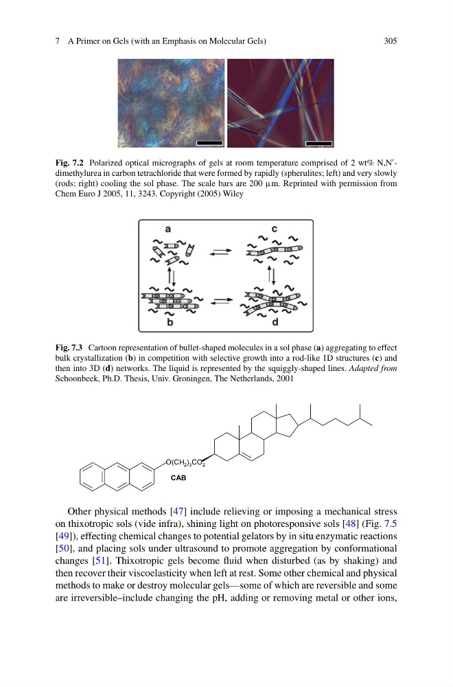
Fig. 7.2 Polarized optical micrographs of gels at room temperature comprised of 2 wt% N,N -
dimethylurea in carbon tetrachloride that were formed by rapidly (spherulites; left) and very slowly
(rods; right) cooling the sol phase. The scale bars are 200 μm. Reprinted with permission from
Chem Euro J 2005, 11, 3243. Copyright (2005) Wiley
Fig. 7.3 Cartoon representation of bullet-shaped molecules in a sol phase (a) aggregating to effect
bulk crystallization (b) in competition with selective growth into a rod-like 1D structures (c) and
then into 3D (d) networks. The liquid is represented by the squiggly-shaped lines. Adapted from
Schoonbeek, Ph.D. Thesis, Univ. Groningen, The Netherlands, 2001
CAB
O(CH 2 ) 3 CO 2
Other physical methods [47] include relieving or imposing a mechanical stress
on thixotropic sols (vide infra), shining light on photoresponsive sols [48] (Fig. 7.5
[49]), effecting chemical changes to potential gelators by in situ enzymatic reactions
[50], and placing sols under ultrasound to promote aggregation by conformational
changes [51]. Thixotropic gels become fluid when disturbed (as by shaking) and
then recover their viscoelasticity when left at rest. Some other chemical and physical
methods to make or destroy molecular gels—some of which are reversible and some
are irreversible–include changing the pH, adding or removing metal or other ions,
305
Fig. 7.2 Polarized optical micrographs of gels at room temperature comprised of 2 wt% N,N -
dimethylurea in carbon tetrachloride that were formed by rapidly (spherulites; left) and very slowly
(rods; right) cooling the sol phase. The scale bars are 200 μm. Reprinted with permission from
Chem Euro J 2005, 11, 3243. Copyright (2005) Wiley
Fig. 7.3 Cartoon representation of bullet-shaped molecules in a sol phase (a) aggregating to effect
bulk crystallization (b) in competition with selective growth into a rod-like 1D structures (c) and
then into 3D (d) networks. The liquid is represented by the squiggly-shaped lines. Adapted from
Schoonbeek, Ph.D. Thesis, Univ. Groningen, The Netherlands, 2001
CAB
O(CH 2 ) 3 CO 2
Other physical methods [47] include relieving or imposing a mechanical stress
on thixotropic sols (vide infra), shining light on photoresponsive sols [48] (Fig. 7.5
[49]), effecting chemical changes to potential gelators by in situ enzymatic reactions
[50], and placing sols under ultrasound to promote aggregation by conformational
changes [51]. Thixotropic gels become fluid when disturbed (as by shaking) and
then recover their viscoelasticity when left at rest. Some other chemical and physical
methods to make or destroy molecular gels—some of which are reversible and some
are irreversible–include changing the pH, adding or removing metal or other ions,