

280
W. Qiu and X.-Y. Liu
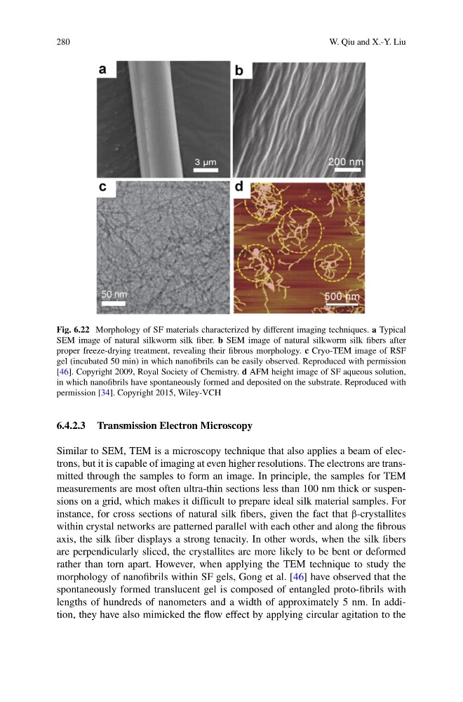
Fig. 6.22 Morphology of SF materials characterized by different imaging techniques. a Typical
SEM image of natural silkworm silk fiber. b SEM image of natural silkworm silk fibers after
proper freeze-drying treatment, revealing their fibrous morphology. c Cryo-TEM image of RSF
gel (incubated 50 min) in which nanofibrils can be easily observed. Reproduced with permission
[46]. Copyright 2009, Royal Society of Chemistry. d AFM height image of SF aqueous solution,
in which nanofibrils have spontaneously formed and deposited on the substrate. Reproduced with
permission [34]. Copyright 2015, Wiley-VCH
6.4.2.3 Transmission Electron Microscopy
Similar to SEM, TEM is a microscopy technique that also applies a beam of electrons, but it is capable of imaging at even higher resolutions. The electrons are transmitted through the samples to form an image. In principle, the samples for TEM
measurements are most often ultra-thin sections less than 100 nm thick or suspensions on a grid, which makes it difficult to prepare ideal silk material samples. For
instance, for cross sections of natural silk fibers, given the fact that β-crystallites
within crystal networks are patterned parallel with each other and along the fibrous
axis, the silk fiber displays a strong tenacity. In other words, when the silk fibers
are perpendicularly sliced, the crystallites are more likely to be bent or deformed
rather than torn apart. However, when applying the TEM technique to study the
morphology of nanofibrils within SF gels, Gong et al. [46] have observed that the
spontaneously formed translucent gel is composed of entangled proto-fibrils with
lengths of hundreds of nanometers and a width of approximately 5 nm. In addition, they have also mimicked the flow effect by applying circular agitation to the
W. Qiu and X.-Y. Liu
Fig. 6.22 Morphology of SF materials characterized by different imaging techniques. a Typical
SEM image of natural silkworm silk fiber. b SEM image of natural silkworm silk fibers after
proper freeze-drying treatment, revealing their fibrous morphology. c Cryo-TEM image of RSF
gel (incubated 50 min) in which nanofibrils can be easily observed. Reproduced with permission
[46]. Copyright 2009, Royal Society of Chemistry. d AFM height image of SF aqueous solution,
in which nanofibrils have spontaneously formed and deposited on the substrate. Reproduced with
permission [34]. Copyright 2015, Wiley-VCH
6.4.2.3 Transmission Electron Microscopy
Similar to SEM, TEM is a microscopy technique that also applies a beam of electrons, but it is capable of imaging at even higher resolutions. The electrons are transmitted through the samples to form an image. In principle, the samples for TEM
measurements are most often ultra-thin sections less than 100 nm thick or suspensions on a grid, which makes it difficult to prepare ideal silk material samples. For
instance, for cross sections of natural silk fibers, given the fact that β-crystallites
within crystal networks are patterned parallel with each other and along the fibrous
axis, the silk fiber displays a strong tenacity. In other words, when the silk fibers
are perpendicularly sliced, the crystallites are more likely to be bent or deformed
rather than torn apart. However, when applying the TEM technique to study the
morphology of nanofibrils within SF gels, Gong et al. [46] have observed that the
spontaneously formed translucent gel is composed of entangled proto-fibrils with
lengths of hundreds of nanometers and a width of approximately 5 nm. In addition, they have also mimicked the flow effect by applying circular agitation to the