

388
C. Thorn and A. Shore
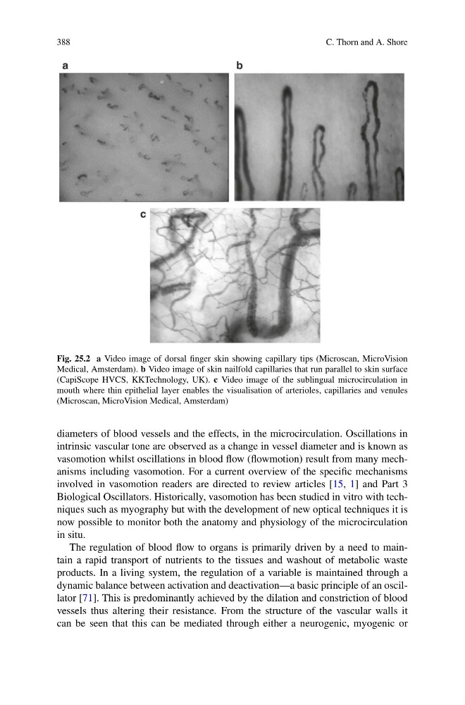
Fig. 25.2 a Video image of dorsal finger skin showing capillary tips (Microscan, MicroVision
Medical, Amsterdam). b Video image of skin nailfold capillaries that run parallel to skin surface
(CapiScope HVCS, KKTechnology, UK). c Video image of the sublingual microcirculation in
mouth where thin epithelial layer enables the visualisation of arterioles, capillaries and venules
(Microscan, MicroVision Medical, Amsterdam)
diameters of blood vessels and the effects, in the microcirculation. Oscillations in
intrinsic vascular tone are observed as a change in vessel diameter and is known as
vasomotion whilst oscillations in blood flow (flowmotion) result from many mechanisms including vasomotion. For a current overview of the specific mechanisms
involved in vasomotion readers are directed to review articles [15, 1] and Part 3
Biological Oscillators. Historically, vasomotion has been studied in vitro with techniques such as myography but with the development of new optical techniques it is
now possible to monitor both the anatomy and physiology of the microcirculation
in situ.
The regulation of blood flow to organs is primarily driven by a need to maintain a rapid transport of nutrients to the tissues and washout of metabolic waste
products. In a living system, the regulation of a variable is maintained through a
dynamic balance between activation and deactivation—a basic principle of an oscillator [71]. This is predominantly achieved by the dilation and constriction of blood
vessels thus altering their resistance. From the structure of the vascular walls it
can be seen that this can be mediated through either a neurogenic, myogenic or
C. Thorn and A. Shore
Fig. 25.2 a Video image of dorsal finger skin showing capillary tips (Microscan, MicroVision
Medical, Amsterdam). b Video image of skin nailfold capillaries that run parallel to skin surface
(CapiScope HVCS, KKTechnology, UK). c Video image of the sublingual microcirculation in
mouth where thin epithelial layer enables the visualisation of arterioles, capillaries and venules
(Microscan, MicroVision Medical, Amsterdam)
diameters of blood vessels and the effects, in the microcirculation. Oscillations in
intrinsic vascular tone are observed as a change in vessel diameter and is known as
vasomotion whilst oscillations in blood flow (flowmotion) result from many mechanisms including vasomotion. For a current overview of the specific mechanisms
involved in vasomotion readers are directed to review articles [15, 1] and Part 3
Biological Oscillators. Historically, vasomotion has been studied in vitro with techniques such as myography but with the development of new optical techniques it is
now possible to monitor both the anatomy and physiology of the microcirculation
in situ.
The regulation of blood flow to organs is primarily driven by a need to maintain a rapid transport of nutrients to the tissues and washout of metabolic waste
products. In a living system, the regulation of a variable is maintained through a
dynamic balance between activation and deactivation—a basic principle of an oscillator [71]. This is predominantly achieved by the dilation and constriction of blood
vessels thus altering their resistance. From the structure of the vascular walls it
can be seen that this can be mediated through either a neurogenic, myogenic or