

21 Brain Morphological and Functional Networks …
335
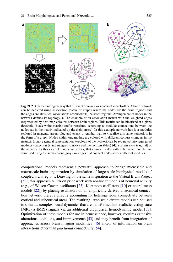
Fig. 21.2 Characterizing the way that different brain regions connect to each other. A brain network
can be depicted using association matrix or graphs where the nodes are the brain regions and
the edges are statistical associations (connections) between regions. Arrangement of nodes in the
network defines its topology. a The example of an association matrix with the weighted edges
(represented by heat-map colours) between brain regions. This matrix can be binarized at a given
threshold (black-white matrix) and/or reordered according to modular connections between the
nodes (as in the matrix indicated by the right arrow). In this example network has four modules
(colored in magenta, green, blue and cyan). b Another way to visualize this same network is in
the form of a graph. Nodes within one module are colored with different colours (same as in the
matrix). In more general representation, topology of the network can be separated into segregated
modules (magenta) (c and integrative nodes and interactions (blue) (d). e Brain view (sagittal) of
the network. In this example nodes and edges, that connect nodes within the same module, are
visuilised using the same colour, grays are edges that connect nodes across different modules
computational models represent a powerful approach to bridge microscale and
macroscale brain organization by simulation of large-scale biophysical models of
coupled brain regions. Drawing on the same inspiration as the Virtual Brain Project
[59], this approach builds on prior work with nonlinear models of neuronal activity
(e.g., of Wilson-Cowan oscillators [23], Kuramoto oscillators [10] or neural mass
models [22]) by placing oscillators on an empirically-derived anatomical connection network, thereby directly accounting for heterogeneous connectivity between
cortical and subcortical areas. The resulting large-scale circuit models can be used
to simulate complex neural dynamics that are transformed into realistic resting-state
fMRI (rs-fMRI) signals via an additional biophysical hemodynamic model [33].
Optimization of these models for use in neuroscience, however, requires extensive
alterations, additions, and improvements [53] and may benefit from integration of
approaches across brain imaging modalities [46] and/or of information on brain
interactions other than functional connectivity [54].
335
Fig. 21.2 Characterizing the way that different brain regions connect to each other. A brain network
can be depicted using association matrix or graphs where the nodes are the brain regions and
the edges are statistical associations (connections) between regions. Arrangement of nodes in the
network defines its topology. a The example of an association matrix with the weighted edges
(represented by heat-map colours) between brain regions. This matrix can be binarized at a given
threshold (black-white matrix) and/or reordered according to modular connections between the
nodes (as in the matrix indicated by the right arrow). In this example network has four modules
(colored in magenta, green, blue and cyan). b Another way to visualize this same network is in
the form of a graph. Nodes within one module are colored with different colours (same as in the
matrix). In more general representation, topology of the network can be separated into segregated
modules (magenta) (c and integrative nodes and interactions (blue) (d). e Brain view (sagittal) of
the network. In this example nodes and edges, that connect nodes within the same module, are
visuilised using the same colour, grays are edges that connect nodes across different modules
computational models represent a powerful approach to bridge microscale and
macroscale brain organization by simulation of large-scale biophysical models of
coupled brain regions. Drawing on the same inspiration as the Virtual Brain Project
[59], this approach builds on prior work with nonlinear models of neuronal activity
(e.g., of Wilson-Cowan oscillators [23], Kuramoto oscillators [10] or neural mass
models [22]) by placing oscillators on an empirically-derived anatomical connection network, thereby directly accounting for heterogeneous connectivity between
cortical and subcortical areas. The resulting large-scale circuit models can be used
to simulate complex neural dynamics that are transformed into realistic resting-state
fMRI (rs-fMRI) signals via an additional biophysical hemodynamic model [33].
Optimization of these models for use in neuroscience, however, requires extensive
alterations, additions, and improvements [53] and may benefit from integration of
approaches across brain imaging modalities [46] and/or of information on brain
interactions other than functional connectivity [54].