

332
V. Vuksanovi´ c
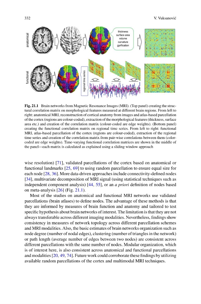
Fig. 21.1 Brain networks from Magnetic Resonance Images (MRI). (Top panel) creating the structural correlation matrix on morphological features measured at different brain regions. From left to
right: anatomical MRI, reconstruction of cortical anatomy from images and atlas-based parcellation
of the cortex (regions are colour-coded), extraction of the morphological features (thickness, surface
area etc.) and creation of the correlation matrix (colour-coded are edge weights). (Bottom panel)
creating the functional correlation matrix on regional time series. From left to right: functional
MRI, atlas-based parcellation of the cortex (regions are colour-coded), extraction of the regional
time series and creation of the correlation matrix from pair-wise correlations between them (colorcoded are edge weights). Time-varying functional correlation matrices are shown in the middle of
the panel—each matrix is calculated as explained using a sliding window approach
wise resolution) [71], validated parcellations of the cortex based on anatomical or
functional landmarks [25, 69] to using random parcellation to ensure equal size for
each node [28, 36]. More data-driven approaches include connectivity-defined nodes
[34], multivariate decomposition of MRI signal (using statistical techniques such as
independent component analysis) [44, 55], or an a priori definition of nodes based
on meta-analysis [26] (Fig. 21.1).
Most of the studies on anatomical and functional MRI networks use validated
parcellations (brain atlases) to define nodes. The advantage of these methods is that
they are informed by measures of brain function and anatomy and tailored to test
specific hypothesis about brain networks of interest. The limitation is that they are not
always transferable across different imaging modalities. Nevertheless, findings show
consistency in measures of network topology across different parcellation schemes
and MRI modalities. Also, the basic estimates of brain networks organization such as
node degree (number of nodal edges), clustering (number of triangles in the network)
or path length (average number of edges between two nodes) are consistent across
different parcellations with the same number of nodes. Modular organization, which
is of interest here, is also consistent across anatomical and functional parcellations
and modalities [20, 49, 74]. Future work could corroborate these findings by utilizing
available random parcellations of the cortex and multimodal MRI techniques.
V. Vuksanovi´ c
Fig. 21.1 Brain networks from Magnetic Resonance Images (MRI). (Top panel) creating the structural correlation matrix on morphological features measured at different brain regions. From left to
right: anatomical MRI, reconstruction of cortical anatomy from images and atlas-based parcellation
of the cortex (regions are colour-coded), extraction of the morphological features (thickness, surface
area etc.) and creation of the correlation matrix (colour-coded are edge weights). (Bottom panel)
creating the functional correlation matrix on regional time series. From left to right: functional
MRI, atlas-based parcellation of the cortex (regions are colour-coded), extraction of the regional
time series and creation of the correlation matrix from pair-wise correlations between them (colorcoded are edge weights). Time-varying functional correlation matrices are shown in the middle of
the panel—each matrix is calculated as explained using a sliding window approach
wise resolution) [71], validated parcellations of the cortex based on anatomical or
functional landmarks [25, 69] to using random parcellation to ensure equal size for
each node [28, 36]. More data-driven approaches include connectivity-defined nodes
[34], multivariate decomposition of MRI signal (using statistical techniques such as
independent component analysis) [44, 55], or an a priori definition of nodes based
on meta-analysis [26] (Fig. 21.1).
Most of the studies on anatomical and functional MRI networks use validated
parcellations (brain atlases) to define nodes. The advantage of these methods is that
they are informed by measures of brain function and anatomy and tailored to test
specific hypothesis about brain networks of interest. The limitation is that they are not
always transferable across different imaging modalities. Nevertheless, findings show
consistency in measures of network topology across different parcellation schemes
and MRI modalities. Also, the basic estimates of brain networks organization such as
node degree (number of nodal edges), clustering (number of triangles in the network)
or path length (average number of edges between two nodes) are consistent across
different parcellations with the same number of nodes. Modular organization, which
is of interest here, is also consistent across anatomical and functional parcellations
and modalities [20, 49, 74]. Future work could corroborate these findings by utilizing
available random parcellations of the cortex and multimodal MRI techniques.