

2
P. R. Willmott
work fn Th
GaN gap
F excimer
2
S
O
N
C
Se
U
m c
e
2
Co
ionization energy
H−atom
Cu Kα
10 eV
1 eV
1 keV
10 keV
100 keV
1 MeV
100 eV
soft x−ray
tender
gamma
hard x−ray
UV
vis
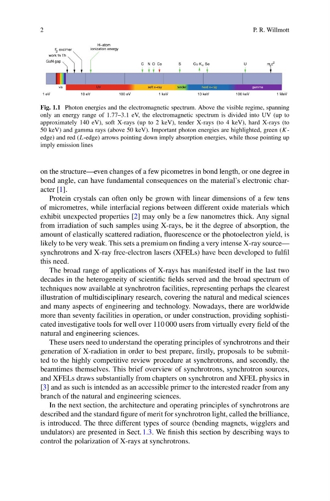
Fig. 1.1 Photon energies and the electromagnetic spectrum. Above the visible regime, spanning
only an energy range of 1.77–3.1 eV, the electromagnetic spectrum is divided into UV (up to
approximately 140 eV), soft X-rays (up to 2 keV), tender X-rays (to 4 keV), hard X-rays (to
50 keV) and gamma rays (above 50 keV). Important photon energies are highlighted, green (K -
edge) and red (L-edge) arrows pointing down imply absorption energies, while those pointing up
imply emission lines
on the structure—even changes of a few picometres in bond length, or one degree in
bond angle, can have fundamental consequences on the material’s electronic character [1].
Protein crystals can often only be grown with linear dimensions of a few tens
of micrometres, while interfacial regions between different oxide materials which
exhibit unexpected properties [2] may only be a few nanometres thick. Any signal
from irradiation of such samples using X-rays, be it the degree of absorption, the
amount of elastically scattered radiation, fluorescence or the photoelectron yield, is
likely to be very weak. This sets a premium on finding a very intense X-ray source—
synchrotrons and X-ray free-electron lasers (XFELs) have been developed to fulfil
this need.
The broad range of applications of X-rays has manifested itself in the last two
decades in the heterogeneity of scientific fields served and the broad spectrum of
techniques now available at synchrotron facilities, representing perhaps the clearest
illustration of multidisciplinary research, covering the natural and medical sciences
and many aspects of engineering and technology. Nowadays, there are worldwide
more than seventy facilities in operation, or under construction, providing sophisticated investigative tools for well over 110 000 users from virtually every field of the
natural and engineering sciences.
These users need to understand the operating principles of synchrotrons and their
generation of X-radiation in order to best prepare, firstly, proposals to be submitted to the highly competitive review procedure at synchrotrons, and secondly, the
beamtimes themselves. This brief overview of synchrotrons, synchrotron sources,
and XFELs draws substantially from chapters on synchrotron and XFEL physics in
[3] and as such is intended as an accessible primer to the interested reader from any
branch of the natural and engineering sciences.
In the next section, the architecture and operating principles of synchrotrons are
described and the standard figure of merit for synchrotron light, called the brilliance,
is introduced. The three different types of source (bending magnets, wigglers and
undulators) are presented in Sect. 1.3. We finish this section by describing ways to
control the polarization of X-rays at synchrotrons.
P. R. Willmott
work fn Th
GaN gap
F excimer
2
S
O
N
C
Se
U
m c
e
2
Co
ionization energy
H−atom
Cu Kα
10 eV
1 eV
1 keV
10 keV
100 keV
1 MeV
100 eV
soft x−ray
tender
gamma
hard x−ray
UV
vis
Fig. 1.1 Photon energies and the electromagnetic spectrum. Above the visible regime, spanning
only an energy range of 1.77–3.1 eV, the electromagnetic spectrum is divided into UV (up to
approximately 140 eV), soft X-rays (up to 2 keV), tender X-rays (to 4 keV), hard X-rays (to
50 keV) and gamma rays (above 50 keV). Important photon energies are highlighted, green (K -
edge) and red (L-edge) arrows pointing down imply absorption energies, while those pointing up
imply emission lines
on the structure—even changes of a few picometres in bond length, or one degree in
bond angle, can have fundamental consequences on the material’s electronic character [1].
Protein crystals can often only be grown with linear dimensions of a few tens
of micrometres, while interfacial regions between different oxide materials which
exhibit unexpected properties [2] may only be a few nanometres thick. Any signal
from irradiation of such samples using X-rays, be it the degree of absorption, the
amount of elastically scattered radiation, fluorescence or the photoelectron yield, is
likely to be very weak. This sets a premium on finding a very intense X-ray source—
synchrotrons and X-ray free-electron lasers (XFELs) have been developed to fulfil
this need.
The broad range of applications of X-rays has manifested itself in the last two
decades in the heterogeneity of scientific fields served and the broad spectrum of
techniques now available at synchrotron facilities, representing perhaps the clearest
illustration of multidisciplinary research, covering the natural and medical sciences
and many aspects of engineering and technology. Nowadays, there are worldwide
more than seventy facilities in operation, or under construction, providing sophisticated investigative tools for well over 110 000 users from virtually every field of the
natural and engineering sciences.
These users need to understand the operating principles of synchrotrons and their
generation of X-radiation in order to best prepare, firstly, proposals to be submitted to the highly competitive review procedure at synchrotrons, and secondly, the
beamtimes themselves. This brief overview of synchrotrons, synchrotron sources,
and XFELs draws substantially from chapters on synchrotron and XFEL physics in
[3] and as such is intended as an accessible primer to the interested reader from any
branch of the natural and engineering sciences.
In the next section, the architecture and operating principles of synchrotrons are
described and the standard figure of merit for synchrotron light, called the brilliance,
is introduced. The three different types of source (bending magnets, wigglers and
undulators) are presented in Sect. 1.3. We finish this section by describing ways to
control the polarization of X-rays at synchrotrons.