

478
T. Balusamy et al.
Oliveira [34]. Roland et al. [23] have investigated the fatigue behavior of untreated
316L stainless steel and those subjected to SMAT. The fatigue strength of the SMAT
316L stainless steel is found to be much higher than the untreated one at both low
and high cycle fatigue regimes. The extent of an increase in fatigue strength for the
treated stainless steel is much pronounced at high cycle fatigue. The surface nanocrystallization and the compressive residual stress induced by SMAT offered considerable resistance for the propagation of fatigue cracks. Post-annealing of SMAT 316L
stainless steels at 400 °C also enhanced fatigue strength. In spite of the relaxation
in compressive residual stress, the increase in surface ductility, limited increase in
grain size, strain hardening and strain-induced martensitic transformation accounts
for the improvement in fatigue strength.
Bahl et al. [24] have evaluated the corrosion fatigue of microcrystalline (MC)
and nanocrystalline (NC) 316L stainless steels in 0.9% NaCl at 0.1 and 5 Hz for up
to 106 cycles or to the point of complete fracture. The stress vs. number of cycles
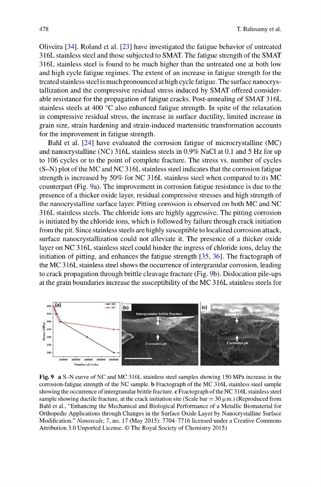
(S–N) plot of the MC and NC 316L stainless steel indicates that the corrosion fatigue
strength is increased by 50% for NC 316L stainless steel when compared to its MC
counterpart (Fig. 9a). The improvement in corrosion fatigue resistance is due to the
presence of a thicker oxide layer, residual compressive stresses and high strength of
the nanocrystalline surface layer. Pitting corrosion is observed on both MC and NC
316L stainless steels. The chloride ions are highly aggressive. The pitting corrosion
is initiated by the chloride ions, which is followed by failure through crack initiation
from the pit. Since stainless steels are highly susceptible to localized corrosion attack,
surface nanocrystallization could not alleviate it. The presence of a thicker oxide
layer on NC 316L stainless steel could hinder the ingress of chloride ions, delay the
initiation of pitting, and enhances the fatigue strength [35, 36]. The fractograph of
the MC 316L stainless steel shows the occurrence of intergranular corrosion, leading
to crack propagation through brittle cleavage fracture (Fig. 9b). Dislocation pile-ups
at the grain boundaries increase the susceptibility of the MC 316L stainless steels for
Fig. 9 a S–N curve of NC and MC 316L stainless steel samples showing 150 MPa increase in the
corrosion-fatigue strength of the NC sample. b Fractograph of the MC 316L stainless steel sample
showing the occurrence of intergranular brittle fracture. c Fractograph of the NC 316L stainless steel
sample showing ductile fracture, at the crack initiation site (Scale bar = 30 μm.) (Reproduced from
Bahl et al., “Enhancing the Mechanical and Biological Performance of a Metallic Biomaterial for
Orthopedic Applications through Changes in the Surface Oxide Layer by Nanocrystalline Surface
Modification.” Nanoscale, 7, no. 17 (May 2015): 7704–7716 licensed under a Creative Commons
Attribution 3.0 Unported License. © The Royal Society of Chemistry 2015)
T. Balusamy et al.
Oliveira [34]. Roland et al. [23] have investigated the fatigue behavior of untreated
316L stainless steel and those subjected to SMAT. The fatigue strength of the SMAT
316L stainless steel is found to be much higher than the untreated one at both low
and high cycle fatigue regimes. The extent of an increase in fatigue strength for the
treated stainless steel is much pronounced at high cycle fatigue. The surface nanocrystallization and the compressive residual stress induced by SMAT offered considerable resistance for the propagation of fatigue cracks. Post-annealing of SMAT 316L
stainless steels at 400 °C also enhanced fatigue strength. In spite of the relaxation
in compressive residual stress, the increase in surface ductility, limited increase in
grain size, strain hardening and strain-induced martensitic transformation accounts
for the improvement in fatigue strength.
Bahl et al. [24] have evaluated the corrosion fatigue of microcrystalline (MC)
and nanocrystalline (NC) 316L stainless steels in 0.9% NaCl at 0.1 and 5 Hz for up
to 106 cycles or to the point of complete fracture. The stress vs. number of cycles
(S–N) plot of the MC and NC 316L stainless steel indicates that the corrosion fatigue
strength is increased by 50% for NC 316L stainless steel when compared to its MC
counterpart (Fig. 9a). The improvement in corrosion fatigue resistance is due to the
presence of a thicker oxide layer, residual compressive stresses and high strength of
the nanocrystalline surface layer. Pitting corrosion is observed on both MC and NC
316L stainless steels. The chloride ions are highly aggressive. The pitting corrosion
is initiated by the chloride ions, which is followed by failure through crack initiation
from the pit. Since stainless steels are highly susceptible to localized corrosion attack,
surface nanocrystallization could not alleviate it. The presence of a thicker oxide
layer on NC 316L stainless steel could hinder the ingress of chloride ions, delay the
initiation of pitting, and enhances the fatigue strength [35, 36]. The fractograph of
the MC 316L stainless steel shows the occurrence of intergranular corrosion, leading
to crack propagation through brittle cleavage fracture (Fig. 9b). Dislocation pile-ups
at the grain boundaries increase the susceptibility of the MC 316L stainless steels for
Fig. 9 a S–N curve of NC and MC 316L stainless steel samples showing 150 MPa increase in the
corrosion-fatigue strength of the NC sample. b Fractograph of the MC 316L stainless steel sample
showing the occurrence of intergranular brittle fracture. c Fractograph of the NC 316L stainless steel
sample showing ductile fracture, at the crack initiation site (Scale bar = 30 μm.) (Reproduced from
Bahl et al., “Enhancing the Mechanical and Biological Performance of a Metallic Biomaterial for
Orthopedic Applications through Changes in the Surface Oxide Layer by Nanocrystalline Surface
Modification.” Nanoscale, 7, no. 17 (May 2015): 7704–7716 licensed under a Creative Commons
Attribution 3.0 Unported License. © The Royal Society of Chemistry 2015)