

Laser Induced Micro/Nano …
455
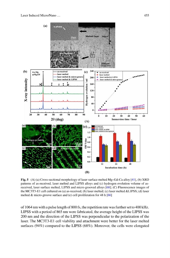
Fig. 5 (A) (a) Cross-sectional morphology of laser surface melted Mg–Gd-Ca alloy [41], (b) XRD
patterns of as-received, laser melted and LIPSS alloys and (c) hydrogen evolution volume of asreceived, laser surface melted, LIPSS and micro-grooved alloys [68]; (C) Fluorescence images of
the MC3T3-E1 cell cultured on (a) as-received; (b) laser melted; (c) laser melted &LIPSS; (d) laser
melted & micro-groove surface and (e) cell proliferation for 48 h [66]
of 1064 nm with a pulse length of 800 fs, the repetition rate was further set to 400 kHz.
LIPSS with a period of 865 nm were fabricated, the average height of the LIPSS was
200 nm and the direction of the LIPSS was perpendicular to the polarization of the
laser. The MC3T3-E1 cell viability and attachment were better for the laser melted
surfaces (94%) compared to the LIPSS (68%). Moreover, the cells were elongated
455
Fig. 5 (A) (a) Cross-sectional morphology of laser surface melted Mg–Gd-Ca alloy [41], (b) XRD
patterns of as-received, laser melted and LIPSS alloys and (c) hydrogen evolution volume of asreceived, laser surface melted, LIPSS and micro-grooved alloys [68]; (C) Fluorescence images of
the MC3T3-E1 cell cultured on (a) as-received; (b) laser melted; (c) laser melted &LIPSS; (d) laser
melted & micro-groove surface and (e) cell proliferation for 48 h [66]
of 1064 nm with a pulse length of 800 fs, the repetition rate was further set to 400 kHz.
LIPSS with a period of 865 nm were fabricated, the average height of the LIPSS was
200 nm and the direction of the LIPSS was perpendicular to the polarization of the
laser. The MC3T3-E1 cell viability and attachment were better for the laser melted
surfaces (94%) compared to the LIPSS (68%). Moreover, the cells were elongated