

376
N. Ashwin Kumar et al.
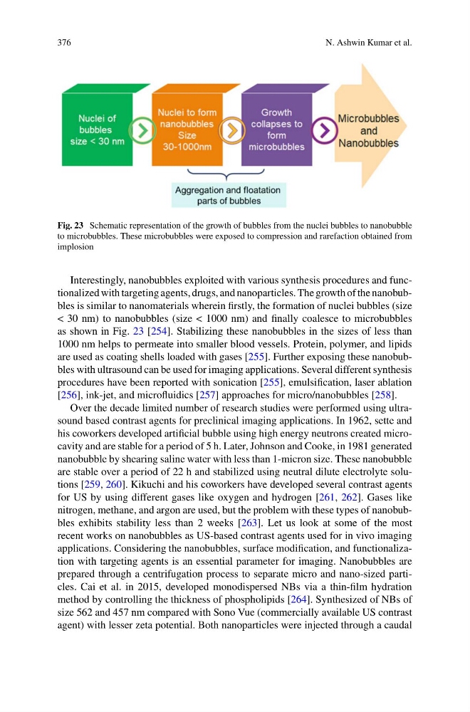
Fig. 23 Schematic representation of the growth of bubbles from the nuclei bubbles to nanobubble
to microbubbles. These microbubbles were exposed to compression and rarefaction obtained from
implosion
Interestingly, nanobubbles exploited with various synthesis procedures and functionalized with targeting agents, drugs, and nanoparticles. The growth of the nanobubbles is similar to nanomaterials wherein firstly, the formation of nuclei bubbles (size
< 30 nm) to nanobubbles (size < 1000 nm) and finally coalesce to microbubbles
as shown in Fig. 23 [254]. Stabilizing these nanobubbles in the sizes of less than
1000 nm helps to permeate into smaller blood vessels. Protein, polymer, and lipids
are used as coating shells loaded with gases [255]. Further exposing these nanobubbles with ultrasound can be used for imaging applications. Several different synthesis
procedures have been reported with sonication [255], emulsification, laser ablation
[256], ink-jet, and microfluidics [257] approaches for micro/nanobubbles [258].
Over the decade limited number of research studies were performed using ultrasound based contrast agents for preclinical imaging applications. In 1962, sette and
his coworkers developed artificial bubble using high energy neutrons created microcavity and are stable for a period of 5 h. Later, Johnson and Cooke, in 1981 generated
nanobubble by shearing saline water with less than 1-micron size. These nanobubble
are stable over a period of 22 h and stabilized using neutral dilute electrolyte solutions [259, 260]. Kikuchi and his coworkers have developed several contrast agents
for US by using different gases like oxygen and hydrogen [261, 262]. Gases like
nitrogen, methane, and argon are used, but the problem with these types of nanobubbles exhibits stability less than 2 weeks [263]. Let us look at some of the most
recent works on nanobubbles as US-based contrast agents used for in vivo imaging
applications. Considering the nanobubbles, surface modification, and functionalization with targeting agents is an essential parameter for imaging. Nanobubbles are
prepared through a centrifugation process to separate micro and nano-sized particles. Cai et al. in 2015, developed monodispersed NBs via a thin-film hydration
method by controlling the thickness of phospholipids [264]. Synthesized of NBs of
size 562 and 457 nm compared with Sono Vue (commercially available US contrast
agent) with lesser zeta potential. Both nanoparticles were injected through a caudal
N. Ashwin Kumar et al.
Fig. 23 Schematic representation of the growth of bubbles from the nuclei bubbles to nanobubble
to microbubbles. These microbubbles were exposed to compression and rarefaction obtained from
implosion
Interestingly, nanobubbles exploited with various synthesis procedures and functionalized with targeting agents, drugs, and nanoparticles. The growth of the nanobubbles is similar to nanomaterials wherein firstly, the formation of nuclei bubbles (size
< 30 nm) to nanobubbles (size < 1000 nm) and finally coalesce to microbubbles
as shown in Fig. 23 [254]. Stabilizing these nanobubbles in the sizes of less than
1000 nm helps to permeate into smaller blood vessels. Protein, polymer, and lipids
are used as coating shells loaded with gases [255]. Further exposing these nanobubbles with ultrasound can be used for imaging applications. Several different synthesis
procedures have been reported with sonication [255], emulsification, laser ablation
[256], ink-jet, and microfluidics [257] approaches for micro/nanobubbles [258].
Over the decade limited number of research studies were performed using ultrasound based contrast agents for preclinical imaging applications. In 1962, sette and
his coworkers developed artificial bubble using high energy neutrons created microcavity and are stable for a period of 5 h. Later, Johnson and Cooke, in 1981 generated
nanobubble by shearing saline water with less than 1-micron size. These nanobubble
are stable over a period of 22 h and stabilized using neutral dilute electrolyte solutions [259, 260]. Kikuchi and his coworkers have developed several contrast agents
for US by using different gases like oxygen and hydrogen [261, 262]. Gases like
nitrogen, methane, and argon are used, but the problem with these types of nanobubbles exhibits stability less than 2 weeks [263]. Let us look at some of the most
recent works on nanobubbles as US-based contrast agents used for in vivo imaging
applications. Considering the nanobubbles, surface modification, and functionalization with targeting agents is an essential parameter for imaging. Nanobubbles are
prepared through a centrifugation process to separate micro and nano-sized particles. Cai et al. in 2015, developed monodispersed NBs via a thin-film hydration
method by controlling the thickness of phospholipids [264]. Synthesized of NBs of
size 562 and 457 nm compared with Sono Vue (commercially available US contrast
agent) with lesser zeta potential. Both nanoparticles were injected through a caudal