

374
N. Ashwin Kumar et al.
7.5 Targeted MNOPs
Targeting molecules attached on the surface of MONPs had an improved MR signal
from the tumor region. Li et al. synthesized PEG capped MONPs as a nano-based
contrast to understand molecular aspects of renal carcinoma using MR imaging [241].
MONPs exposed with 3 T MRI, PEG-MONPs showed (12.94 mM s
−1 ) three times
higher relaxivity than gadolinium chelates. Aptamer (AS1411), a potential target that
recognizes nucleolin and the ability to internalize quickly with renal carcinoma. MR
images were acquired for 7 days determines the higher contrast in tumor, liver, and
kidneys at 45 min of post-injection. PEG-MONPs were cleared in 24 h compared
to AS1411-MONPs, wherein clearance within 7 days via renal filtration. Nano theranostics agents were developed using MONPs functionalized with polydopamine
(melanin pigment as photothermal agent) linked with folic acid to localize particles into the cancer regime. Ding and his coworkers triggered MONPs with NIR to
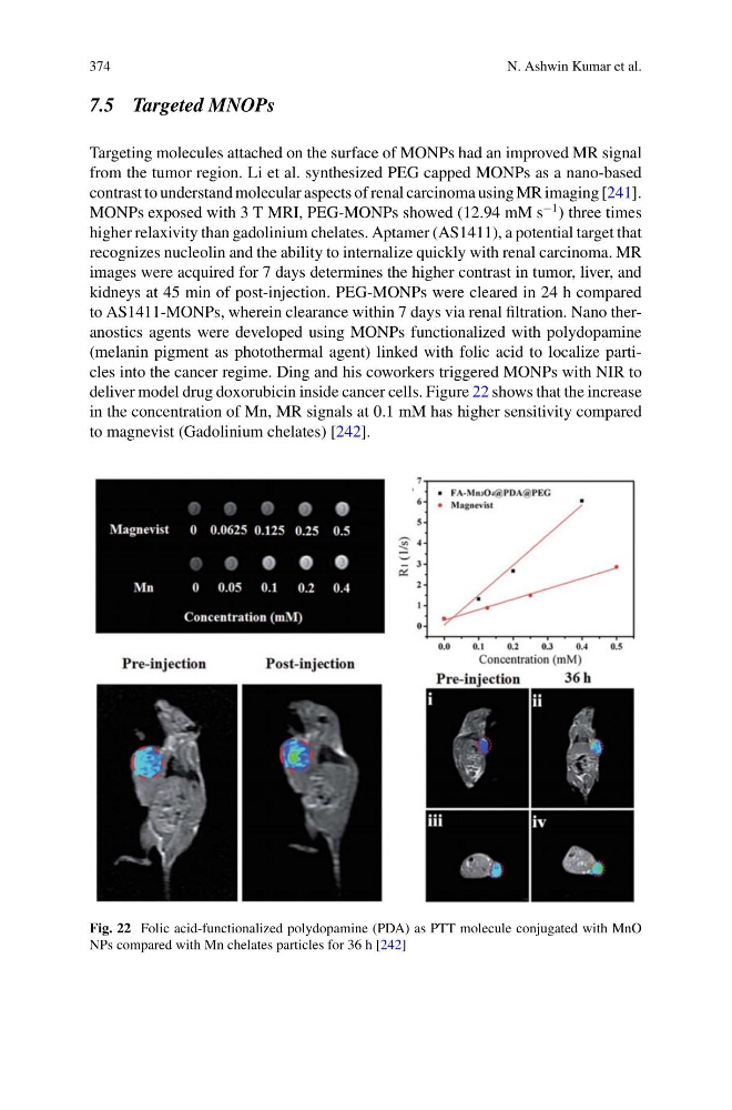
deliver model drug doxorubicin inside cancer cells. Figure 22 shows that the increase
in the concentration of Mn, MR signals at 0.1 mM has higher sensitivity compared
to magnevist (Gadolinium chelates) [242].
Fig. 22 Folic acid-functionalized polydopamine (PDA) as PTT molecule conjugated with MnO
NPs compared with Mn chelates particles for 36 h [242]
N. Ashwin Kumar et al.
7.5 Targeted MNOPs
Targeting molecules attached on the surface of MONPs had an improved MR signal
from the tumor region. Li et al. synthesized PEG capped MONPs as a nano-based
contrast to understand molecular aspects of renal carcinoma using MR imaging [241].
MONPs exposed with 3 T MRI, PEG-MONPs showed (12.94 mM s
−1 ) three times
higher relaxivity than gadolinium chelates. Aptamer (AS1411), a potential target that
recognizes nucleolin and the ability to internalize quickly with renal carcinoma. MR
images were acquired for 7 days determines the higher contrast in tumor, liver, and
kidneys at 45 min of post-injection. PEG-MONPs were cleared in 24 h compared
to AS1411-MONPs, wherein clearance within 7 days via renal filtration. Nano theranostics agents were developed using MONPs functionalized with polydopamine
(melanin pigment as photothermal agent) linked with folic acid to localize particles into the cancer regime. Ding and his coworkers triggered MONPs with NIR to
deliver model drug doxorubicin inside cancer cells. Figure 22 shows that the increase
in the concentration of Mn, MR signals at 0.1 mM has higher sensitivity compared
to magnevist (Gadolinium chelates) [242].
Fig. 22 Folic acid-functionalized polydopamine (PDA) as PTT molecule conjugated with MnO
NPs compared with Mn chelates particles for 36 h [242]