

362
N. Ashwin Kumar et al.
disadvantage of MRI is the contrast between the tissues within the organs is negligible. For instance, the delineation of the tumor region and the normal tissues show
a poor diagnosis for physicians. This separation between the normal and abnormal
regions requires a localized exogenous contrast to detect via micro-MRI is necessary [148]. Exogenous contrast agents are an excellent supplement for the current
medical imaging systems to enhance the diagnosis. New types of nanomaterials are
being developed to improve the contrast between the tissues in both single as well
as multi-modal imaging aspects [149]. Paramagnetic (T1 contrast agents) and superparamagnetic (T2 iron oxide nanoparticles) properties were used in MRI to acquire
the images and to enhance the contrast between the tissues [150].
7.1 Gadolinium Chelates Based Contrast Agents
Species of Gadolinium ions were chelated with different compounds that are used as
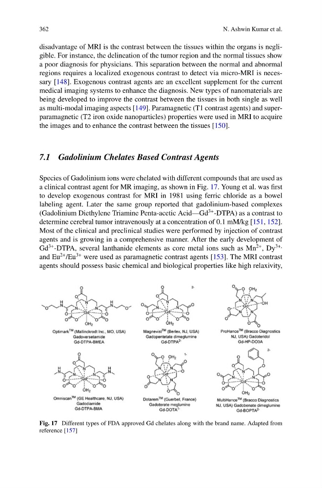
a clinical contrast agent for MR imaging, as shown in Fig. 17. Young et al. was first
to develop exogenous contrast for MRI in 1981 using ferric chloride as a bowel
labeling agent. Later the same group reported that gadolinium-based complexes
(Gadolinium Diethylene Triamine Penta-acetic Acid—Gd
3+ -DTPA) as a contrast to
determine cerebral tumor intravenously at a concentration of 0.1 mM/kg [151, 152].
Most of the clinical and preclinical studies were performed by injection of contrast
agents and is growing in a comprehensive manner. After the early development of
Gd
3+ -DTPA, several lanthanide elements as core metal ions such as Mn
2+ , Dy
3+,
and Eu
2+ /Eu
3+ were used as paramagnetic contrast agents [153]. The MRI contrast
agents should possess basic chemical and biological properties like high relaxivity,
Fig. 17 Different types of FDA approved Gd chelates along with the brand name. Adapted from
reference [157]
N. Ashwin Kumar et al.
disadvantage of MRI is the contrast between the tissues within the organs is negligible. For instance, the delineation of the tumor region and the normal tissues show
a poor diagnosis for physicians. This separation between the normal and abnormal
regions requires a localized exogenous contrast to detect via micro-MRI is necessary [148]. Exogenous contrast agents are an excellent supplement for the current
medical imaging systems to enhance the diagnosis. New types of nanomaterials are
being developed to improve the contrast between the tissues in both single as well
as multi-modal imaging aspects [149]. Paramagnetic (T1 contrast agents) and superparamagnetic (T2 iron oxide nanoparticles) properties were used in MRI to acquire
the images and to enhance the contrast between the tissues [150].
7.1 Gadolinium Chelates Based Contrast Agents
Species of Gadolinium ions were chelated with different compounds that are used as
a clinical contrast agent for MR imaging, as shown in Fig. 17. Young et al. was first
to develop exogenous contrast for MRI in 1981 using ferric chloride as a bowel
labeling agent. Later the same group reported that gadolinium-based complexes
(Gadolinium Diethylene Triamine Penta-acetic Acid—Gd
3+ -DTPA) as a contrast to
determine cerebral tumor intravenously at a concentration of 0.1 mM/kg [151, 152].
Most of the clinical and preclinical studies were performed by injection of contrast
agents and is growing in a comprehensive manner. After the early development of
Gd
3+ -DTPA, several lanthanide elements as core metal ions such as Mn
2+ , Dy
3+,
and Eu
2+ /Eu
3+ were used as paramagnetic contrast agents [153]. The MRI contrast
agents should possess basic chemical and biological properties like high relaxivity,
Fig. 17 Different types of FDA approved Gd chelates along with the brand name. Adapted from
reference [157]