

Nanomaterials for Medical Imaging …
349
glioblastoma, and leukemia would overexpress similar markers in humans and in vivo
[79, 80]. In this context, a simple approach made by Zhou et al. conjugating GNPs
with folic acid using a branched poly ethylenimine and coated with PEG [81]. They
produced water-soluble GNPs with a size around 2.1 nm, which is non-toxic, and
pre-clinical CT imaging was performed at different time points. Due to PEG functionalized on GNPs, they were not considered as foreign bodies and started to accumulate
in heart, kidney, tumor, and other regions. Further circulation for 72 h in vivo, there
is an increased GNPs accumulation compared to a previous time point. Eck et al.,
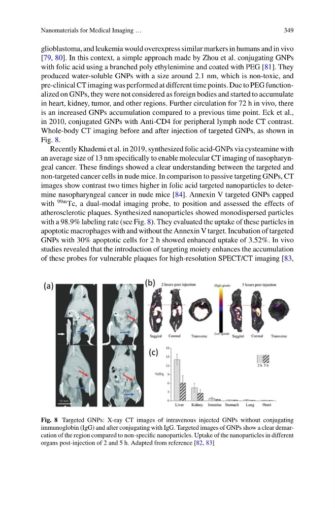
in 2010, conjugated GNPs with Anti-CD4 for peripheral lymph node CT contrast.
Whole-body CT imaging before and after injection of targeted GNPs, as shown in
Fig. 8.
Recently Khademi et al. in 2019, synthesized folic acid-GNPs via cysteamine with
an average size of 13 nm specifically to enable molecular CT imaging of nasopharyngeal cancer. These findings showed a clear understanding between the targeted and
non-targeted cancer cells in nude mice. In comparison to passive targeting GNPs, CT
images show contrast two times higher in folic acid targeted nanoparticles to determine nasopharyngeal cancer in nude mice [84]. Annexin V targeted GNPs capped
with
99m Tc, a dual-modal imaging probe, to position and assessed the effects of
atherosclerotic plaques. Synthesized nanoparticles showed monodispersed particles
with a 98.9% labeling rate (see Fig. 8). They evaluated the uptake of these particles in
apoptotic macrophages with and without the Annexin V target. Incubation of targeted
GNPs with 30% apoptotic cells for 2 h showed enhanced uptake of 3.52%. In vivo
studies revealed that the introduction of targeting moiety enhances the accumulation
of these probes for vulnerable plaques for high-resolution SPECT/CT imaging [83,
Fig. 8 Targeted GNPs: X-ray CT images of intravenous injected GNPs without conjugating
immunoglobin (IgG) and after conjugating with IgG. Targeted images of GNPs show a clear demarcation of the region compared to non-specific nanoparticles. Uptake of the nanoparticles in different
organs post-injection of 2 and 5 h. Adapted from reference [82, 83]
349
glioblastoma, and leukemia would overexpress similar markers in humans and in vivo
[79, 80]. In this context, a simple approach made by Zhou et al. conjugating GNPs
with folic acid using a branched poly ethylenimine and coated with PEG [81]. They
produced water-soluble GNPs with a size around 2.1 nm, which is non-toxic, and
pre-clinical CT imaging was performed at different time points. Due to PEG functionalized on GNPs, they were not considered as foreign bodies and started to accumulate
in heart, kidney, tumor, and other regions. Further circulation for 72 h in vivo, there
is an increased GNPs accumulation compared to a previous time point. Eck et al.,
in 2010, conjugated GNPs with Anti-CD4 for peripheral lymph node CT contrast.
Whole-body CT imaging before and after injection of targeted GNPs, as shown in
Fig. 8.
Recently Khademi et al. in 2019, synthesized folic acid-GNPs via cysteamine with
an average size of 13 nm specifically to enable molecular CT imaging of nasopharyngeal cancer. These findings showed a clear understanding between the targeted and
non-targeted cancer cells in nude mice. In comparison to passive targeting GNPs, CT
images show contrast two times higher in folic acid targeted nanoparticles to determine nasopharyngeal cancer in nude mice [84]. Annexin V targeted GNPs capped
with
99m Tc, a dual-modal imaging probe, to position and assessed the effects of
atherosclerotic plaques. Synthesized nanoparticles showed monodispersed particles
with a 98.9% labeling rate (see Fig. 8). They evaluated the uptake of these particles in
apoptotic macrophages with and without the Annexin V target. Incubation of targeted
GNPs with 30% apoptotic cells for 2 h showed enhanced uptake of 3.52%. In vivo
studies revealed that the introduction of targeting moiety enhances the accumulation
of these probes for vulnerable plaques for high-resolution SPECT/CT imaging [83,
Fig. 8 Targeted GNPs: X-ray CT images of intravenous injected GNPs without conjugating
immunoglobin (IgG) and after conjugating with IgG. Targeted images of GNPs show a clear demarcation of the region compared to non-specific nanoparticles. Uptake of the nanoparticles in different
organs post-injection of 2 and 5 h. Adapted from reference [82, 83]