

184
S. T. Aruna and M. Shilpa
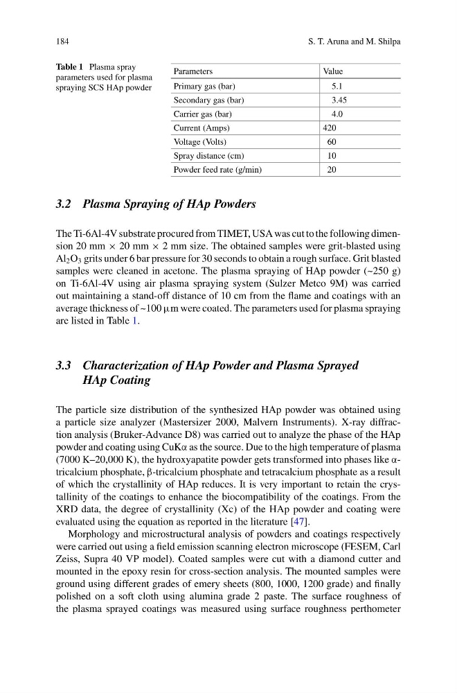
Table 1 Plasma spray
parameters used for plasma
spraying SCS HAp powder
Parameters
Value
Primary gas (bar)
5.1
Secondary gas (bar)
3.45
Carrier gas (bar)
4.0
Current (Amps)
420
Voltage (Volts)
60
Spray distance (cm)
10
Powder feed rate (g/min)
20
3.2 Plasma Spraying of HAp Powders
The Ti-6Al-4V substrate procured from TIMET, USA was cut to the following dimension 20 mm × 20 mm × 2 mm size. The obtained samples were grit-blasted using
Al 2 O 3 grits under 6 bar pressure for 30 seconds to obtain a rough surface. Grit blasted
samples were cleaned in acetone. The plasma spraying of HAp powder (~250 g)
on Ti-6Al-4V using air plasma spraying system (Sulzer Metco 9M) was carried
out maintaining a stand-off distance of 10 cm from the flame and coatings with an
average thickness of ~100 μm were coated. The parameters used for plasma spraying
are listed in Table 1.
3.3 Characterization of HAp Powder and Plasma Sprayed
HAp Coating
The particle size distribution of the synthesized HAp powder was obtained using
a particle size analyzer (Mastersizer 2000, Malvern Instruments). X-ray diffraction analysis (Bruker-Advance D8) was carried out to analyze the phase of the HAp
powder and coating using CuKα as the source. Due to the high temperature of plasma
(7000 K–20,000 K), the hydroxyapatite powder gets transformed into phases like αtricalcium phosphate, β-tricalcium phosphate and tetracalcium phosphate as a result
of which the crystallinity of HAp reduces. It is very important to retain the crystallinity of the coatings to enhance the biocompatibility of the coatings. From the
XRD data, the degree of crystallinity (Xc) of the HAp powder and coating were
evaluated using the equation as reported in the literature [47].
Morphology and microstructural analysis of powders and coatings respectively
were carried out using a field emission scanning electron microscope (FESEM, Carl
Zeiss, Supra 40 VP model). Coated samples were cut with a diamond cutter and
mounted in the epoxy resin for cross-section analysis. The mounted samples were
ground using different grades of emery sheets (800, 1000, 1200 grade) and finally
polished on a soft cloth using alumina grade 2 paste. The surface roughness of
the plasma sprayed coatings was measured using surface roughness perthometer
S. T. Aruna and M. Shilpa
Table 1 Plasma spray
parameters used for plasma
spraying SCS HAp powder
Parameters
Value
Primary gas (bar)
5.1
Secondary gas (bar)
3.45
Carrier gas (bar)
4.0
Current (Amps)
420
Voltage (Volts)
60
Spray distance (cm)
10
Powder feed rate (g/min)
20
3.2 Plasma Spraying of HAp Powders
The Ti-6Al-4V substrate procured from TIMET, USA was cut to the following dimension 20 mm × 20 mm × 2 mm size. The obtained samples were grit-blasted using
Al 2 O 3 grits under 6 bar pressure for 30 seconds to obtain a rough surface. Grit blasted
samples were cleaned in acetone. The plasma spraying of HAp powder (~250 g)
on Ti-6Al-4V using air plasma spraying system (Sulzer Metco 9M) was carried
out maintaining a stand-off distance of 10 cm from the flame and coatings with an
average thickness of ~100 μm were coated. The parameters used for plasma spraying
are listed in Table 1.
3.3 Characterization of HAp Powder and Plasma Sprayed
HAp Coating
The particle size distribution of the synthesized HAp powder was obtained using
a particle size analyzer (Mastersizer 2000, Malvern Instruments). X-ray diffraction analysis (Bruker-Advance D8) was carried out to analyze the phase of the HAp
powder and coating using CuKα as the source. Due to the high temperature of plasma
(7000 K–20,000 K), the hydroxyapatite powder gets transformed into phases like αtricalcium phosphate, β-tricalcium phosphate and tetracalcium phosphate as a result
of which the crystallinity of HAp reduces. It is very important to retain the crystallinity of the coatings to enhance the biocompatibility of the coatings. From the
XRD data, the degree of crystallinity (Xc) of the HAp powder and coating were
evaluated using the equation as reported in the literature [47].
Morphology and microstructural analysis of powders and coatings respectively
were carried out using a field emission scanning electron microscope (FESEM, Carl
Zeiss, Supra 40 VP model). Coated samples were cut with a diamond cutter and
mounted in the epoxy resin for cross-section analysis. The mounted samples were
ground using different grades of emery sheets (800, 1000, 1200 grade) and finally
polished on a soft cloth using alumina grade 2 paste. The surface roughness of
the plasma sprayed coatings was measured using surface roughness perthometer