

110
P. Hameed et al.
This chapter is split into two parts comprising the nanofibers as the first part and
nanosurfaces as a second. It helps explain how these processes can help in the overall
osseointegration and antibacterial activity of the biomaterials developed.
2 Part I: Nanofibers
Nanofibers can steer cell alignment [7, 8], enhance cell-cell interaction [9] and
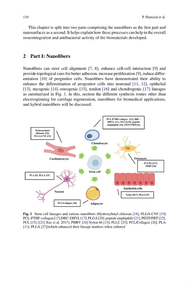
provide topological cues for better adhesion, increase proliferation [9], induce differentiation [10] of progenitor cells. Nanofibers have demonstrated their ability to
enhance the differentiation of progenitor cells into neuronal [11, 12], epithelial
[13], myogenic [14] osteogenic [15], tendon [16] and chondrogenic [17] lineages
as summarized in Fig. 1. In this, section the different synthesis routes other than
electrospinning for cartilage regeneration, nanofibers for biomedical applications,
and hybrid nanofibers will be discussed.
PCL-PTHF-collagen [11]; DBC -
DHYL [11]; PLGA[14]; peptide
amphiphile [15], PEOT/PBT[16]
Hydroxybutyl
chitosan [12];
PLGA-CNF [13]
PLA [5], PLLA [21]
PCL/Collagen [20]
Nylon 66[7]; PLLC[19]
PCL[9] ([17];
PHBV[18]
Fig. 1 Stem cell lineages and various nanofibers (Hydroxybutyl chitosan [18]; PLGA-CNF [19]
PCL-PTHF-collagen [17];DBC-DHYL [17]; PLGA [20]; peptide amphiphile [21], PEOT/PBT [22];
PCL [15] ([23] Xue et al. 2017); PHBV [24] Nylon 66 [13]; PLLC [25]; PCL/Collagen [26]; PLA
[11], PLLA [27])which enhanced their lineage markers when cultured
P. Hameed et al.
This chapter is split into two parts comprising the nanofibers as the first part and
nanosurfaces as a second. It helps explain how these processes can help in the overall
osseointegration and antibacterial activity of the biomaterials developed.
2 Part I: Nanofibers
Nanofibers can steer cell alignment [7, 8], enhance cell-cell interaction [9] and
provide topological cues for better adhesion, increase proliferation [9], induce differentiation [10] of progenitor cells. Nanofibers have demonstrated their ability to
enhance the differentiation of progenitor cells into neuronal [11, 12], epithelial
[13], myogenic [14] osteogenic [15], tendon [16] and chondrogenic [17] lineages
as summarized in Fig. 1. In this, section the different synthesis routes other than
electrospinning for cartilage regeneration, nanofibers for biomedical applications,
and hybrid nanofibers will be discussed.
PCL-PTHF-collagen [11]; DBC -
DHYL [11]; PLGA[14]; peptide
amphiphile [15], PEOT/PBT[16]
Hydroxybutyl
chitosan [12];
PLGA-CNF [13]
PLA [5], PLLA [21]
PCL/Collagen [20]
Nylon 66[7]; PLLC[19]
PCL[9] ([17];
PHBV[18]
Fig. 1 Stem cell lineages and various nanofibers (Hydroxybutyl chitosan [18]; PLGA-CNF [19]
PCL-PTHF-collagen [17];DBC-DHYL [17]; PLGA [20]; peptide amphiphile [21], PEOT/PBT [22];
PCL [15] ([23] Xue et al. 2017); PHBV [24] Nylon 66 [13]; PLLC [25]; PCL/Collagen [26]; PLA
[11], PLLA [27])which enhanced their lineage markers when cultured