

Exploring Non-covalent Interactions by Jet-Cooled Electronic …
73
in the IR spectrum presented in Fig. 6Ba arise due to the N-H…N hydrogen-bonded
N-H stretching of the indole moiety and the free N-H group of the imidazole unit
of the dimer, respectively. The indole…imidazole complex is found to be similar
to the indole…pyridine complex in terms of its structure and the interplay between
different non-covalent interactions present there [115]. Additionally, the structural
motif of the indole…imidazole complex observed from the experiment mimics the
aromatic-aromatic interaction between the side chains of the tryptophan and histidine
residues of proteins [175–177].
Indole…(pyrrole) 2 trimeric complex studied by Kumar et al. represents aromatic
trimeric interactions present in the aromatic side-chains of the amino acid residues
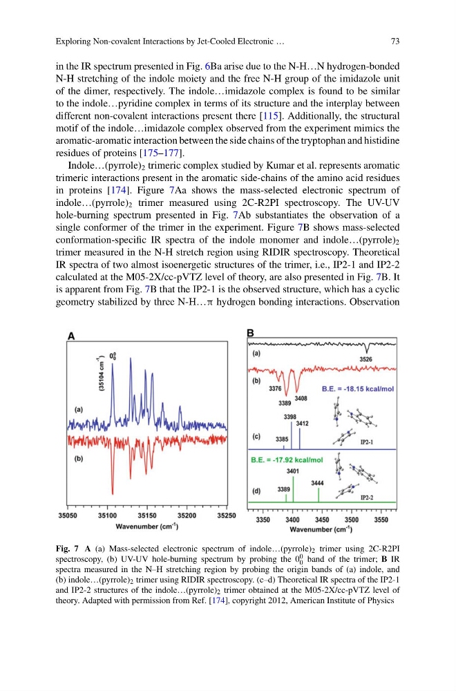
in proteins [174]. Figure 7Aa shows the mass-selected electronic spectrum of
indole…(pyrrole) 2 trimer measured using 2C-R2PI spectroscopy. The UV-UV
hole-burning spectrum presented in Fig. 7Ab substantiates the observation of a
single conformer of the trimer in the experiment. Figure 7B shows mass-selected
conformation-specific IR spectra of the indole monomer and indole…(pyrrole) 2
trimer measured in the N-H stretch region using RIDIR spectroscopy. Theoretical
IR spectra of two almost isoenergetic structures of the trimer, i.e., IP2-1 and IP2-2
calculated at the M05-2X/cc-pVTZ level of theory, are also presented in Fig. 7B. It
is apparent from Fig. 7B that the IP2-1 is the observed structure, which has a cyclic
geometry stabilized by three N-H…π hydrogen bonding interactions. Observation
Fig. 7 A (a) Mass-selected electronic spectrum of indole…(pyrrole) 2 trimer using 2C-R2PI
spectroscopy, (b) UV-UV hole-burning spectrum by probing the 0 0
0 band of the trimer; B IR
spectra measured in the N–H stretching region by probing the origin bands of (a) indole, and
(b) indole…(pyrrole) 2 trimer using RIDIR spectroscopy. (c–d) Theoretical IR spectra of the IP2-1
and IP2-2 structures of the indole…(pyrrole) 2 trimer obtained at the M05-2X/cc-pVTZ level of
theory. Adapted with permission from Ref. [174], copyright 2012, American Institute of Physics
73
in the IR spectrum presented in Fig. 6Ba arise due to the N-H…N hydrogen-bonded
N-H stretching of the indole moiety and the free N-H group of the imidazole unit
of the dimer, respectively. The indole…imidazole complex is found to be similar
to the indole…pyridine complex in terms of its structure and the interplay between
different non-covalent interactions present there [115]. Additionally, the structural
motif of the indole…imidazole complex observed from the experiment mimics the
aromatic-aromatic interaction between the side chains of the tryptophan and histidine
residues of proteins [175–177].
Indole…(pyrrole) 2 trimeric complex studied by Kumar et al. represents aromatic
trimeric interactions present in the aromatic side-chains of the amino acid residues
in proteins [174]. Figure 7Aa shows the mass-selected electronic spectrum of
indole…(pyrrole) 2 trimer measured using 2C-R2PI spectroscopy. The UV-UV
hole-burning spectrum presented in Fig. 7Ab substantiates the observation of a
single conformer of the trimer in the experiment. Figure 7B shows mass-selected
conformation-specific IR spectra of the indole monomer and indole…(pyrrole) 2
trimer measured in the N-H stretch region using RIDIR spectroscopy. Theoretical
IR spectra of two almost isoenergetic structures of the trimer, i.e., IP2-1 and IP2-2
calculated at the M05-2X/cc-pVTZ level of theory, are also presented in Fig. 7B. It
is apparent from Fig. 7B that the IP2-1 is the observed structure, which has a cyclic
geometry stabilized by three N-H…π hydrogen bonding interactions. Observation
Fig. 7 A (a) Mass-selected electronic spectrum of indole…(pyrrole) 2 trimer using 2C-R2PI
spectroscopy, (b) UV-UV hole-burning spectrum by probing the 0 0
0 band of the trimer; B IR
spectra measured in the N–H stretching region by probing the origin bands of (a) indole, and
(b) indole…(pyrrole) 2 trimer using RIDIR spectroscopy. (c–d) Theoretical IR spectra of the IP2-1
and IP2-2 structures of the indole…(pyrrole) 2 trimer obtained at the M05-2X/cc-pVTZ level of
theory. Adapted with permission from Ref. [174], copyright 2012, American Institute of Physics