

Exploring Non-covalent Interactions by Jet-Cooled Electronic …
71
comparison of the experimental and theoretical IR spectra presented in Fig. 5A indicates that the species A and B observed in the experiment are due to indole…pyridine dimer and (indole) 2 …pyridine trimer, respectively. The IR-UV hole-burning
spectra presented in Fig. 5B further confirm that all the electronic bands of the
species A and species B are due to a single conformation of the indole…pyridine
dimer and (indole) 2 …pyridine trimer, respectively. The electronic origin band of
the (indole) 2 …pyridine trimer (A
0
0 ) is red-shifted by 76 cm
−1 while the same of the
indole…pyridine dimer is red-shifted by 271 cm
−1 .
Theoretical calculations performed using wave function theory (MP2) as well as
density functional theory (M05-2X, M06-2X) with different basis sets predict that
the observed indole…pyridine dimer has a V-shaped structure stabilized primarily
by a strong N-H…N hydrogen bond in addition to a secondary C-H…π and weak
π-stacking interactions (Fig. 5Ab). The IR spectrum presented in Fig. 5Ab shows
that the frequency of the N-H…N hydrogen-bonded N-H group of indole in the
indole…pyridine dimer is red-shifted by 257 cm
−1 . The most intriguing finding
from the spectroscopic study of the indole…pyridine dimer is the observation of the
structure which is stabilized due to the interplay between the electrostatic dominated
strong N-H…N hydrogen bond, and dispersion dominated relatively weaker C-H…π
and π-stacking interactions. This result demonstrates that a delicate balance between
the strong and weak non-covalent interactions, which optimize the final shape of
the molecular systems, is the key to the functional structures of biomolecules and
materials.
Energy decomposition analysis of the observed V-shaped structure of the
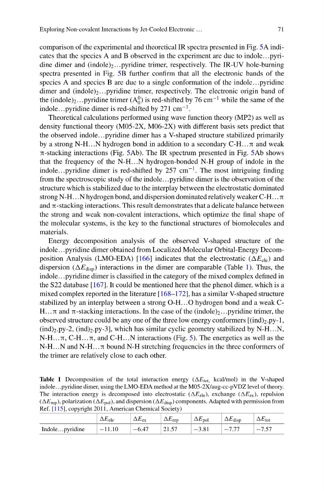
indole…pyridine dimer obtained from Localized Molecular Orbital-Energy Decomposition Analysis (LMO-EDA) [166] indicates that the electrostatic (E ele ) and
dispersion (E disp ) interactions in the dimer are comparable (Table 1). Thus, the
indole…pyridine dimer is classified in the category of the mixed complex defined in
the S22 database [167]. It could be mentioned here that the phenol dimer, which is a
mixed complex reported in the literature [168–172], has a similar V-shaped structure
stabilized by an interplay between a strong O-H…O hydrogen bond and a weak CH…π and π-stacking interactions. In the case of the (indole) 2 …pyridine trimer, the
observed structure could be any one of the three low energy conformers [(ind) 2 .py-1,
(ind) 2 .py-2, (ind) 2 .py-3], which has similar cyclic geometry stabilized by N-H…N,
N-H…π, C-H…π, and C-H…N interactions (Fig. 5). The energetics as well as the
N-H…N and N-H…π bound N-H stretching frequencies in the three conformers of
the trimer are relatively close to each other.
Table 1 Decomposition of the total interaction energy (E tot, kcal/mol) in the V-shaped
indole…pyridine dimer, using the LMO-EDA method at the M05-2X/aug-cc-pVDZ level of theory.
The interaction energy is decomposed into electrostatic (E ele ), exchange (E ex ), repulsion
(E rep ), polarization (E pol ), and dispersion (E disp ) components. Adapted with permission from
Ref. [115], copyright 2011, American Chemical Society)
E ele
E ex
E rep
E pol
E disp
E tot
Indole…pyridine
−11.10
−6.47
21.57
−3.81
−7.77
−7.57
71
comparison of the experimental and theoretical IR spectra presented in Fig. 5A indicates that the species A and B observed in the experiment are due to indole…pyridine dimer and (indole) 2 …pyridine trimer, respectively. The IR-UV hole-burning
spectra presented in Fig. 5B further confirm that all the electronic bands of the
species A and species B are due to a single conformation of the indole…pyridine
dimer and (indole) 2 …pyridine trimer, respectively. The electronic origin band of
the (indole) 2 …pyridine trimer (A
0
0 ) is red-shifted by 76 cm
−1 while the same of the
indole…pyridine dimer is red-shifted by 271 cm
−1 .
Theoretical calculations performed using wave function theory (MP2) as well as
density functional theory (M05-2X, M06-2X) with different basis sets predict that
the observed indole…pyridine dimer has a V-shaped structure stabilized primarily
by a strong N-H…N hydrogen bond in addition to a secondary C-H…π and weak
π-stacking interactions (Fig. 5Ab). The IR spectrum presented in Fig. 5Ab shows
that the frequency of the N-H…N hydrogen-bonded N-H group of indole in the
indole…pyridine dimer is red-shifted by 257 cm
−1 . The most intriguing finding
from the spectroscopic study of the indole…pyridine dimer is the observation of the
structure which is stabilized due to the interplay between the electrostatic dominated
strong N-H…N hydrogen bond, and dispersion dominated relatively weaker C-H…π
and π-stacking interactions. This result demonstrates that a delicate balance between
the strong and weak non-covalent interactions, which optimize the final shape of
the molecular systems, is the key to the functional structures of biomolecules and
materials.
Energy decomposition analysis of the observed V-shaped structure of the
indole…pyridine dimer obtained from Localized Molecular Orbital-Energy Decomposition Analysis (LMO-EDA) [166] indicates that the electrostatic (E ele ) and
dispersion (E disp ) interactions in the dimer are comparable (Table 1). Thus, the
indole…pyridine dimer is classified in the category of the mixed complex defined in
the S22 database [167]. It could be mentioned here that the phenol dimer, which is a
mixed complex reported in the literature [168–172], has a similar V-shaped structure
stabilized by an interplay between a strong O-H…O hydrogen bond and a weak CH…π and π-stacking interactions. In the case of the (indole) 2 …pyridine trimer, the
observed structure could be any one of the three low energy conformers [(ind) 2 .py-1,
(ind) 2 .py-2, (ind) 2 .py-3], which has similar cyclic geometry stabilized by N-H…N,
N-H…π, C-H…π, and C-H…N interactions (Fig. 5). The energetics as well as the
N-H…N and N-H…π bound N-H stretching frequencies in the three conformers of
the trimer are relatively close to each other.
Table 1 Decomposition of the total interaction energy (E tot, kcal/mol) in the V-shaped
indole…pyridine dimer, using the LMO-EDA method at the M05-2X/aug-cc-pVDZ level of theory.
The interaction energy is decomposed into electrostatic (E ele ), exchange (E ex ), repulsion
(E rep ), polarization (E pol ), and dispersion (E disp ) components. Adapted with permission from
Ref. [115], copyright 2011, American Chemical Society)
E ele
E ex
E rep
E pol
E disp
E tot
Indole…pyridine
−11.10
−6.47
21.57
−3.81
−7.77
−7.57