

656
S. P. Singh and S. Siddhanta
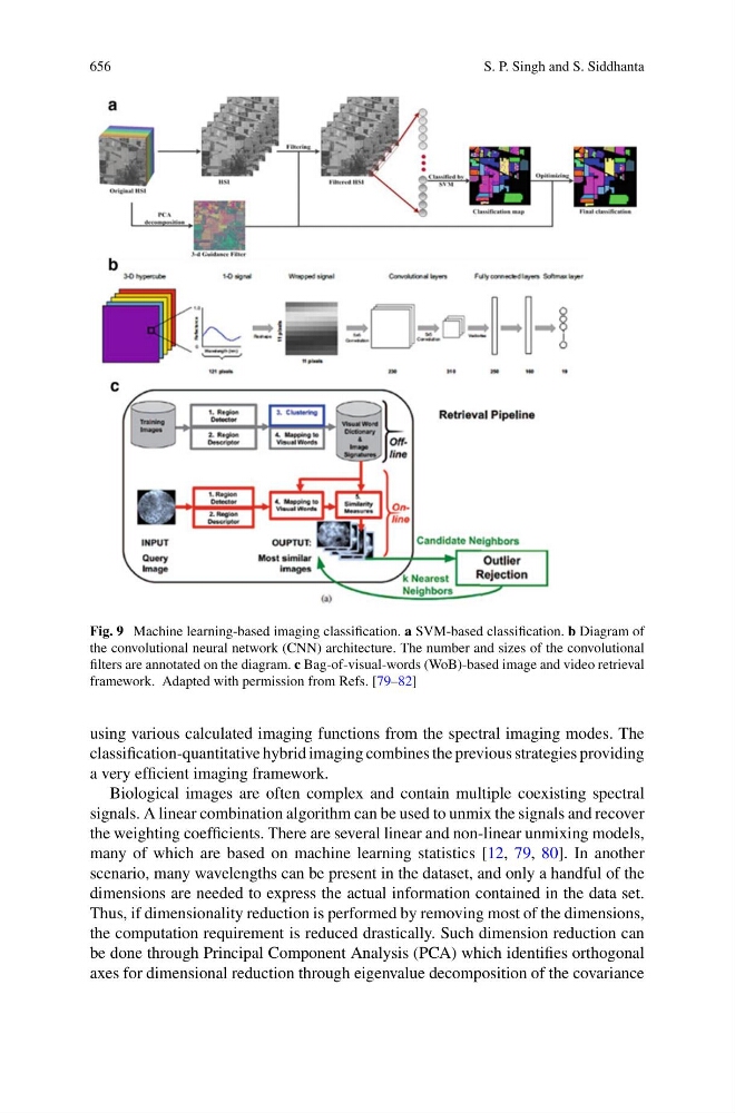
Fig. 9 Machine learning-based imaging classification. a SVM-based classification. b Diagram of
the convolutional neural network (CNN) architecture. The number and sizes of the convolutional
filters are annotated on the diagram. c Bag-of-visual-words (WoB)-based image and video retrieval
framework. Adapted with permission from Refs. [79–82]
using various calculated imaging functions from the spectral imaging modes. The
classification-quantitative hybrid imaging combines the previous strategies providing
a very efficient imaging framework.
Biological images are often complex and contain multiple coexisting spectral
signals. A linear combination algorithm can be used to unmix the signals and recover
the weighting coefficients. There are several linear and non-linear unmixing models,
many of which are based on machine learning statistics [12, 79, 80]. In another
scenario, many wavelengths can be present in the dataset, and only a handful of the
dimensions are needed to express the actual information contained in the data set.
Thus, if dimensionality reduction is performed by removing most of the dimensions,
the computation requirement is reduced drastically. Such dimension reduction can
be done through Principal Component Analysis (PCA) which identifies orthogonal
axes for dimensional reduction through eigenvalue decomposition of the covariance