

Nuclear Magnetic Resonance Spectroscopy: Theory and Applications
507
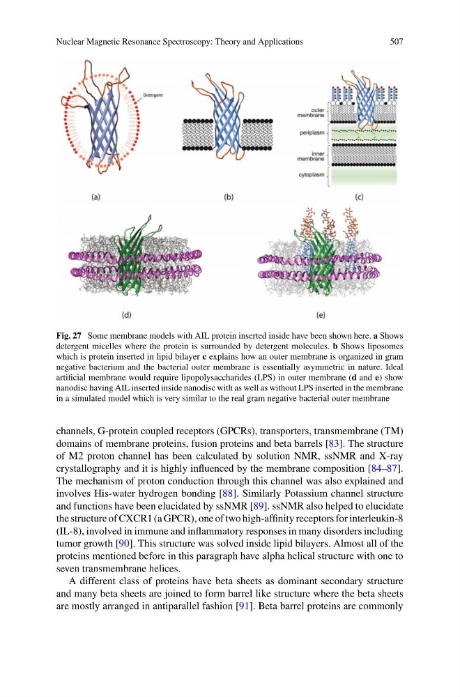
Fig. 27 Some membrane models with AIL protein inserted inside have been shown here. a Shows
detergent micelles where the protein is surrounded by detergent molecules. b Shows liposomes
which is protein inserted in lipid bilayer c explains how an outer membrane is organized in gram
negative bacterium and the bacterial outer membrane is essentially asymmetric in nature. Ideal
artificial membrane would require lipopolysaccharides (LPS) in outer membrane (d and e) show
nanodisc having AIL inserted inside nanodisc with as well as without LPS inserted in the membrane
in a simulated model which is very similar to the real gram negative bacterial outer membrane
channels, G-protein coupled receptors (GPCRs), transporters, transmembrane (TM)
domains of membrane proteins, fusion proteins and beta barrels [83]. The structure
of M2 proton channel has been calculated by solution NMR, ssNMR and X-ray
crystallography and it is highly influenced by the membrane composition [84–87].
The mechanism of proton conduction through this channel was also explained and
involves His-water hydrogen bonding [88]. Similarly Potassium channel structure
and functions have been elucidated by ssNMR [89]. ssNMR also helped to elucidate
the structure of CXCR1 (a GPCR), one of two high-affinity receptors for interleukin-8
(IL-8), involved in immune and inflammatory responses in many disorders including
tumor growth [90]. This structure was solved inside lipid bilayers. Almost all of the
proteins mentioned before in this paragraph have alpha helical structure with one to
seven transmembrane helices.
A different class of proteins have beta sheets as dominant secondary structure
and many beta sheets are joined to form barrel like structure where the beta sheets
are mostly arranged in antiparallel fashion [91]. Beta barrel proteins are commonly
507
Fig. 27 Some membrane models with AIL protein inserted inside have been shown here. a Shows
detergent micelles where the protein is surrounded by detergent molecules. b Shows liposomes
which is protein inserted in lipid bilayer c explains how an outer membrane is organized in gram
negative bacterium and the bacterial outer membrane is essentially asymmetric in nature. Ideal
artificial membrane would require lipopolysaccharides (LPS) in outer membrane (d and e) show
nanodisc having AIL inserted inside nanodisc with as well as without LPS inserted in the membrane
in a simulated model which is very similar to the real gram negative bacterial outer membrane
channels, G-protein coupled receptors (GPCRs), transporters, transmembrane (TM)
domains of membrane proteins, fusion proteins and beta barrels [83]. The structure
of M2 proton channel has been calculated by solution NMR, ssNMR and X-ray
crystallography and it is highly influenced by the membrane composition [84–87].
The mechanism of proton conduction through this channel was also explained and
involves His-water hydrogen bonding [88]. Similarly Potassium channel structure
and functions have been elucidated by ssNMR [89]. ssNMR also helped to elucidate
the structure of CXCR1 (a GPCR), one of two high-affinity receptors for interleukin-8
(IL-8), involved in immune and inflammatory responses in many disorders including
tumor growth [90]. This structure was solved inside lipid bilayers. Almost all of the
proteins mentioned before in this paragraph have alpha helical structure with one to
seven transmembrane helices.
A different class of proteins have beta sheets as dominant secondary structure
and many beta sheets are joined to form barrel like structure where the beta sheets
are mostly arranged in antiparallel fashion [91]. Beta barrel proteins are commonly