

Application of X-Ray Photoelectron Spectroscopy in Materials …
415
example lanthanum hexaboride (LaB 6 ) filament). The energy of the electron
source determines the flux of X-rays produced from the anode.
For XPS, usually Aluminium metal is used as anode and generate X-Rays
with energy 1486.6 eV. These are used as primary source for generating XRay photons. Other than Al, magnesium is also used as anode with energy
1253.6 eV. The energy of the anode decides the maximum binding energy that can
be measured. Moreover, with higher energy of anode material, photoelectrons
with higher kinetic energy will be emitted. Higher energy of the photoelectrons
can be utilised to perform depth analysis in a better way.
3. Monochromator: The X-Rays pass through monochromator, so that only Kα
waves can reach the sample. In addition, these also help in focussing the X-Ray
beam on the sample. As shown in Fig. 1b, the monochromator in XPS is torsoid
in shape, due to which radius of curvature in each direction is different and thus
it become focussed and monochromated. The monochromated X-Rays reach the
sample. The X-Ray spot size on the sample is controlled by the electron beam
spot size on Al anode. Quartz crystal is usually employed as the monochromator.
It follows the Bragg’s equation of diffraction (nλ = 2d sinθ) to focus X-Rays
on the sample. The sample, Al anode and quartz crystal are usually arranged
in a “Rowland circle” of radius r, with quartz crystal in curved shape [1]. The
X-Ray beam pass through a column consisting of a set of lenses (condenser and
objective lens) and alignment coils, which further help in focussing the beam.
4. Analyzer: XPS employ a hemispherical analyser with multichannel detector. The
hemispherical analyzer consist of concentric hemispherical electrodes, wherein
the outer most is the most negative and innermost is most positive. The electrons
pass through the gap in between the concentric electrodes and create the polarity
among the electrodes. As the photoelectrons emitted from the sample are high in
energy and can destroy the detector, they are decelerated using a series of lenses.
Electrons can enter into the analyzer to reach the detector, if they possess kinetic
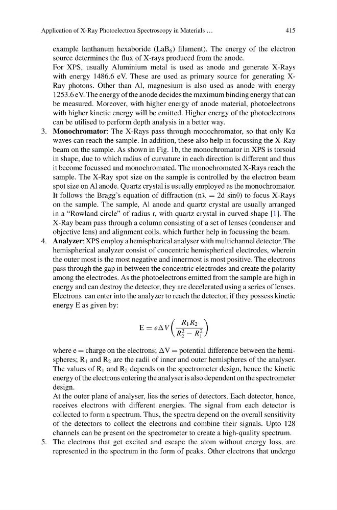
energy E as given by:
E = eV
R 1 R 2
R
2
2 − R
2
1
where e = charge on the electrons; V = potential difference between the hemispheres; R 1 and R 2 are the radii of inner and outer hemispheres of the analyser.
The values of R 1 and R 2 depends on the spectrometer design, hence the kinetic
energy of the electrons entering the analyser is also dependent on the spectrometer
design.
At the outer plane of analyser, lies the series of detectors. Each detector, hence,
receives electrons with different energies. The signal from each detector is
collected to form a spectrum. Thus, the spectra depend on the overall sensitivity
of the detectors to collect the electrons and combine their signals. Upto 128
channels can be present on the spectrometer to create a high-quality spectrum.
5. The electrons that get excited and escape the atom without energy loss, are
represented in the spectrum in the form of peaks. Other electrons that undergo
415
example lanthanum hexaboride (LaB 6 ) filament). The energy of the electron
source determines the flux of X-rays produced from the anode.
For XPS, usually Aluminium metal is used as anode and generate X-Rays
with energy 1486.6 eV. These are used as primary source for generating XRay photons. Other than Al, magnesium is also used as anode with energy
1253.6 eV. The energy of the anode decides the maximum binding energy that can
be measured. Moreover, with higher energy of anode material, photoelectrons
with higher kinetic energy will be emitted. Higher energy of the photoelectrons
can be utilised to perform depth analysis in a better way.
3. Monochromator: The X-Rays pass through monochromator, so that only Kα
waves can reach the sample. In addition, these also help in focussing the X-Ray
beam on the sample. As shown in Fig. 1b, the monochromator in XPS is torsoid
in shape, due to which radius of curvature in each direction is different and thus
it become focussed and monochromated. The monochromated X-Rays reach the
sample. The X-Ray spot size on the sample is controlled by the electron beam
spot size on Al anode. Quartz crystal is usually employed as the monochromator.
It follows the Bragg’s equation of diffraction (nλ = 2d sinθ) to focus X-Rays
on the sample. The sample, Al anode and quartz crystal are usually arranged
in a “Rowland circle” of radius r, with quartz crystal in curved shape [1]. The
X-Ray beam pass through a column consisting of a set of lenses (condenser and
objective lens) and alignment coils, which further help in focussing the beam.
4. Analyzer: XPS employ a hemispherical analyser with multichannel detector. The
hemispherical analyzer consist of concentric hemispherical electrodes, wherein
the outer most is the most negative and innermost is most positive. The electrons
pass through the gap in between the concentric electrodes and create the polarity
among the electrodes. As the photoelectrons emitted from the sample are high in
energy and can destroy the detector, they are decelerated using a series of lenses.
Electrons can enter into the analyzer to reach the detector, if they possess kinetic
energy E as given by:
E = eV
R 1 R 2
R
2
2 − R
2
1
where e = charge on the electrons; V = potential difference between the hemispheres; R 1 and R 2 are the radii of inner and outer hemispheres of the analyser.
The values of R 1 and R 2 depends on the spectrometer design, hence the kinetic
energy of the electrons entering the analyser is also dependent on the spectrometer
design.
At the outer plane of analyser, lies the series of detectors. Each detector, hence,
receives electrons with different energies. The signal from each detector is
collected to form a spectrum. Thus, the spectra depend on the overall sensitivity
of the detectors to collect the electrons and combine their signals. Upto 128
channels can be present on the spectrometer to create a high-quality spectrum.
5. The electrons that get excited and escape the atom without energy loss, are
represented in the spectrum in the form of peaks. Other electrons that undergo