

Modern Experimental Techniques in Ultrafast Atomic …
271
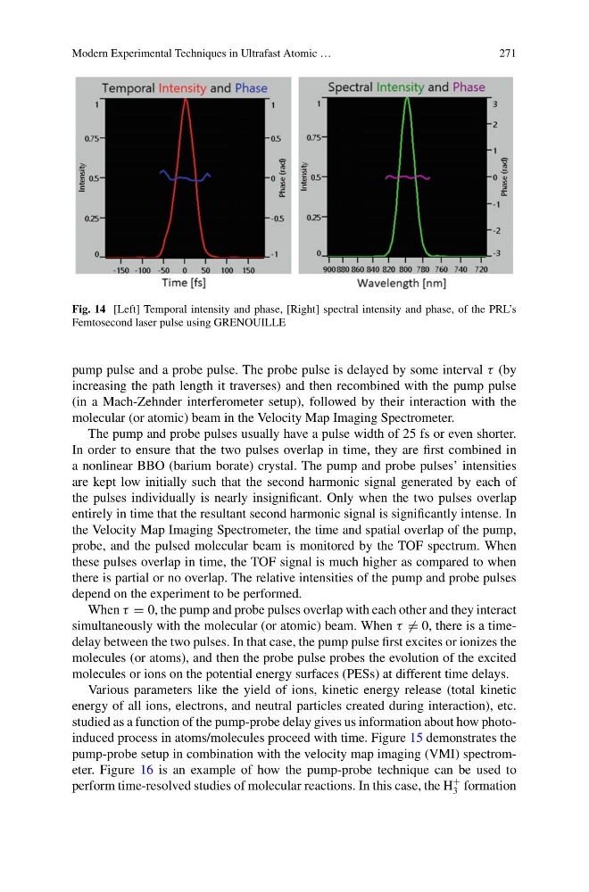
Fig. 14 [Left] Temporal intensity and phase, [Right] spectral intensity and phase, of the PRL’s
Femtosecond laser pulse using GRENOUILLE
pump pulse and a probe pulse. The probe pulse is delayed by some interval τ (by
increasing the path length it traverses) and then recombined with the pump pulse
(in a Mach-Zehnder interferometer setup), followed by their interaction with the
molecular (or atomic) beam in the Velocity Map Imaging Spectrometer.
The pump and probe pulses usually have a pulse width of 25 fs or even shorter.
In order to ensure that the two pulses overlap in time, they are first combined in
a nonlinear BBO (barium borate) crystal. The pump and probe pulses’ intensities
are kept low initially such that the second harmonic signal generated by each of
the pulses individually is nearly insignificant. Only when the two pulses overlap
entirely in time that the resultant second harmonic signal is significantly intense. In
the Velocity Map Imaging Spectrometer, the time and spatial overlap of the pump,
probe, and the pulsed molecular beam is monitored by the TOF spectrum. When
these pulses overlap in time, the TOF signal is much higher as compared to when
there is partial or no overlap. The relative intensities of the pump and probe pulses
depend on the experiment to be performed.
When τ = 0, the pump and probe pulses overlap with each other and they interact
simultaneously with the molecular (or atomic) beam. When τ = 0, there is a timedelay between the two pulses. In that case, the pump pulse first excites or ionizes the
molecules (or atoms), and then the probe pulse probes the evolution of the excited
molecules or ions on the potential energy surfaces (PESs) at different time delays.
Various parameters like the yield of ions, kinetic energy release (total kinetic
energy of all ions, electrons, and neutral particles created during interaction), etc.
studied as a function of the pump-probe delay gives us information about how photoinduced process in atoms/molecules proceed with time. Figure 15 demonstrates the
pump-probe setup in combination with the velocity map imaging (VMI) spectrometer. Figure 16 is an example of how the pump-probe technique can be used to
perform time-resolved studies of molecular reactions. In this case, the H
+
3 formation
271
Fig. 14 [Left] Temporal intensity and phase, [Right] spectral intensity and phase, of the PRL’s
Femtosecond laser pulse using GRENOUILLE
pump pulse and a probe pulse. The probe pulse is delayed by some interval τ (by
increasing the path length it traverses) and then recombined with the pump pulse
(in a Mach-Zehnder interferometer setup), followed by their interaction with the
molecular (or atomic) beam in the Velocity Map Imaging Spectrometer.
The pump and probe pulses usually have a pulse width of 25 fs or even shorter.
In order to ensure that the two pulses overlap in time, they are first combined in
a nonlinear BBO (barium borate) crystal. The pump and probe pulses’ intensities
are kept low initially such that the second harmonic signal generated by each of
the pulses individually is nearly insignificant. Only when the two pulses overlap
entirely in time that the resultant second harmonic signal is significantly intense. In
the Velocity Map Imaging Spectrometer, the time and spatial overlap of the pump,
probe, and the pulsed molecular beam is monitored by the TOF spectrum. When
these pulses overlap in time, the TOF signal is much higher as compared to when
there is partial or no overlap. The relative intensities of the pump and probe pulses
depend on the experiment to be performed.
When τ = 0, the pump and probe pulses overlap with each other and they interact
simultaneously with the molecular (or atomic) beam. When τ = 0, there is a timedelay between the two pulses. In that case, the pump pulse first excites or ionizes the
molecules (or atoms), and then the probe pulse probes the evolution of the excited
molecules or ions on the potential energy surfaces (PESs) at different time delays.
Various parameters like the yield of ions, kinetic energy release (total kinetic
energy of all ions, electrons, and neutral particles created during interaction), etc.
studied as a function of the pump-probe delay gives us information about how photoinduced process in atoms/molecules proceed with time. Figure 15 demonstrates the
pump-probe setup in combination with the velocity map imaging (VMI) spectrometer. Figure 16 is an example of how the pump-probe technique can be used to
perform time-resolved studies of molecular reactions. In this case, the H
+
3 formation