

diffraction patterns were obtained. Changes in basal spacing of layer silicates were
recorded after solvation of the Mg
2+ -saturated samples with glycerin and heating of
the K
+
-saturated samples at 300 and 550
C for 1 h. The clay mineral composition
was evaluated from the basal-spacing changes of the layered silicates (Harris and
White 2008).
The element concentrations in digested or extracted solutions were determined by
colorimetric spectroscopy, atomic absorption spectroscopy, and inductively coupled
plasma spectroscopy. Radiocesium concentrations in soil were determined by
gamma-ray spectrometry (ORTEC, GEM, and DSPEC jr 2.0, 2000s). The radioactivity of soil mineral particles and cross sections of muddy tsunami deposits embedded in resin were detected by an imaging plate (BAS-5000, FUJIFILM Co., Ltd.).
Synchrotron radiation reveals the chemical forms of elements by X-ray absorption near edge structure (XANES) and extended X-ray absorption fine structure
(EXAFS) (Kelly et al. 2008). Originally employed for heavy-element analysis,
XANES and EXAFS are increasingly being applied to lighter elements. Highresolution X-ray ptychography (accurate to 10 nm), X-ray computed tomography,
XANES and EXAFS are expected to become the methods of choice in future
analyses of lighter elements (Ajiboye et al. 2008; Trinh et al. 2017) in soil-plant
systems.
K
L
M
Kα 1
Kβ 1
Lα 1
0
2
4
6
8
Energy (keV)
C
O
Na
Mg
Al
Si
P
Cl
S
K
Ca
Ti V Cr
Mn
Fe
b
a
e -
Characteristic
X-ray
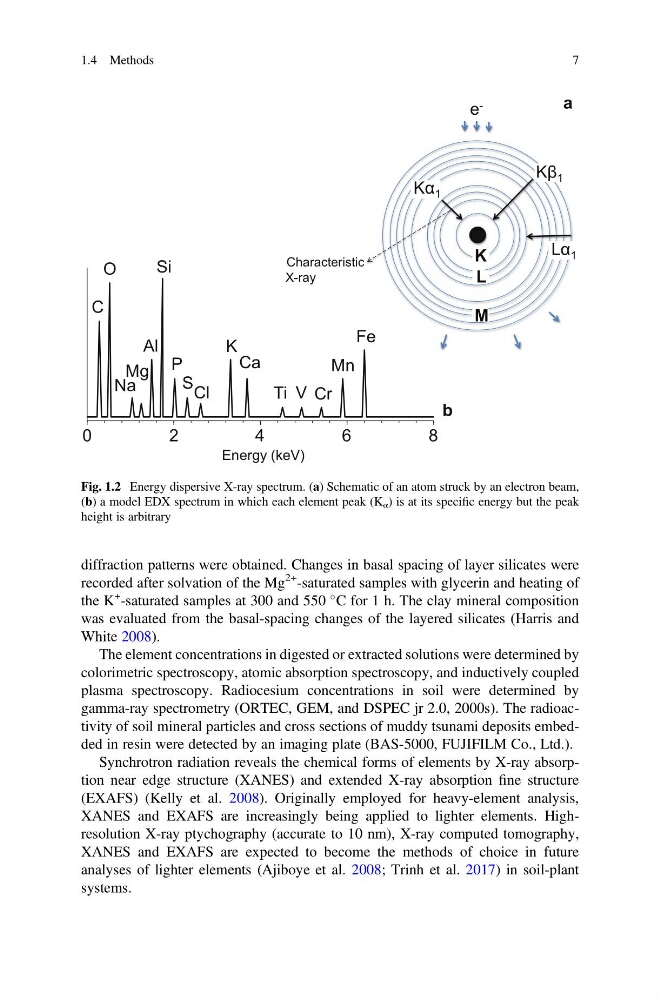
Fig. 1.2 Energy dispersive X-ray spectrum. (a) Schematic of an atom struck by an electron beam,
(b) a model EDX spectrum in which each element peak (K α ) is at its specific energy but the peak
height is arbitrary
1.4 Methods
7
recorded after solvation of the Mg
2+ -saturated samples with glycerin and heating of
the K
+
-saturated samples at 300 and 550
C for 1 h. The clay mineral composition
was evaluated from the basal-spacing changes of the layered silicates (Harris and
White 2008).
The element concentrations in digested or extracted solutions were determined by
colorimetric spectroscopy, atomic absorption spectroscopy, and inductively coupled
plasma spectroscopy. Radiocesium concentrations in soil were determined by
gamma-ray spectrometry (ORTEC, GEM, and DSPEC jr 2.0, 2000s). The radioactivity of soil mineral particles and cross sections of muddy tsunami deposits embedded in resin were detected by an imaging plate (BAS-5000, FUJIFILM Co., Ltd.).
Synchrotron radiation reveals the chemical forms of elements by X-ray absorption near edge structure (XANES) and extended X-ray absorption fine structure
(EXAFS) (Kelly et al. 2008). Originally employed for heavy-element analysis,
XANES and EXAFS are increasingly being applied to lighter elements. Highresolution X-ray ptychography (accurate to 10 nm), X-ray computed tomography,
XANES and EXAFS are expected to become the methods of choice in future
analyses of lighter elements (Ajiboye et al. 2008; Trinh et al. 2017) in soil-plant
systems.
K
L
M
Kα 1
Kβ 1
Lα 1
0
2
4
6
8
Energy (keV)
C
O
Na
Mg
Al
Si
P
Cl
S
K
Ca
Ti V Cr
Mn
Fe
b
a
e -
Characteristic
X-ray
Fig. 1.2 Energy dispersive X-ray spectrum. (a) Schematic of an atom struck by an electron beam,
(b) a model EDX spectrum in which each element peak (K α ) is at its specific energy but the peak
height is arbitrary
1.4 Methods
7