

5.3.4 Vivianite Formation in Bulk Soil
Figures 5.11 and 5.12 showed vivianite formation on rice roots. An advantage of
using rice roots for vivianite detection is the simplicity of separating vivianite from
soil. In addition, iron plaque around rice roots was the site of P accumulation under
reducing soil conditions, which is preferable for vivianite formation, as discussed
above. Incidentally, the occurrence of vivianite crystals on the rice roots suggests
that parts of them are growing from exodermis-like cells toward the bulk soil. It
appears that vivianite can be formed in bulk soil (Zelibor et al. 1988). In fact,
vivianite crystals form aggregates larger than 0.05 mm in diameter. By using soil
with a particle size of less than 0.038 mm, vivianite crystal aggregates can be
separated from the fine soil fraction using a 0.053 mm sieve.
Figure 5.20a shows vivianite crystal aggregates separated from the plow layer soil
of a paddy field under submergence after incubation for 30 days at 30
C. Vivianite
crystals are colorless immediately after separation from the incubated soil. Subsequently, the color of the vivianite crystals slowly turns blue (Fig. 5.20b) by partial
oxidation of Fe
2+ in air (Garnd and Lavkulich 1980). Since other soil minerals are
included in the crystal aggregates, the vivianite content of these aggregates is
approximately 40%.
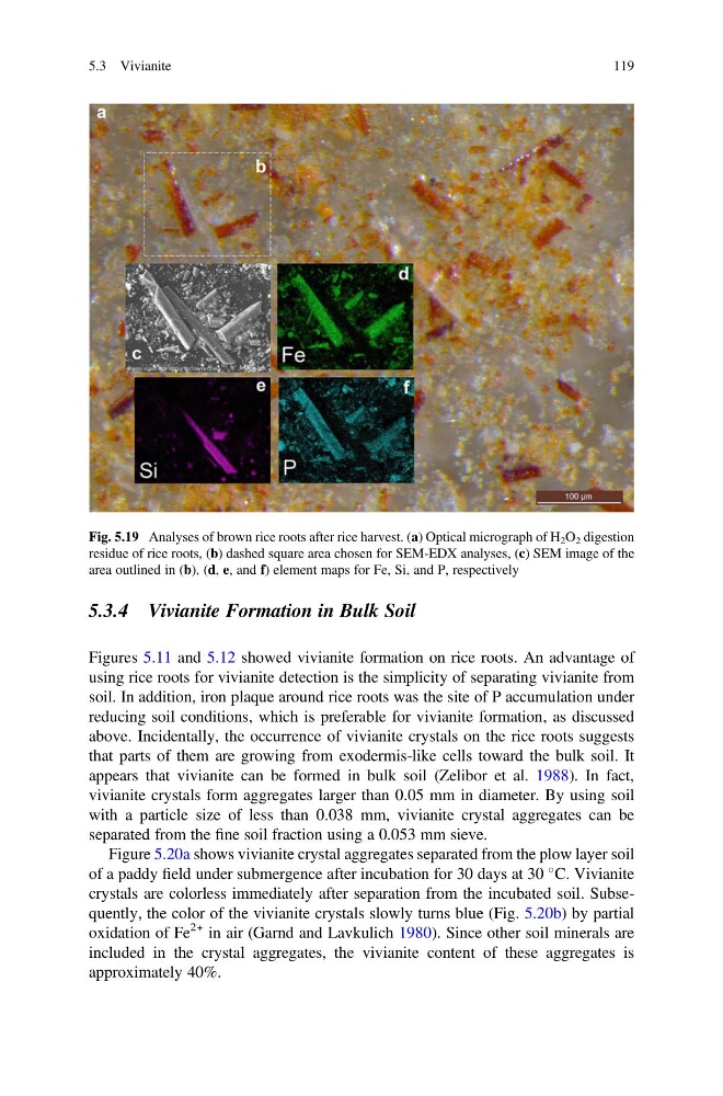
Fig. 5.19 Analyses of brown rice roots after rice harvest. (a) Optical micrograph of H 2 O 2 digestion
residue of rice roots, (b) dashed square area chosen for SEM-EDX analyses, (c) SEM image of the
area outlined in (b), (d, e, and f) element maps for Fe, Si, and P, respectively
5.3 Vivianite
119
Figures 5.11 and 5.12 showed vivianite formation on rice roots. An advantage of
using rice roots for vivianite detection is the simplicity of separating vivianite from
soil. In addition, iron plaque around rice roots was the site of P accumulation under
reducing soil conditions, which is preferable for vivianite formation, as discussed
above. Incidentally, the occurrence of vivianite crystals on the rice roots suggests
that parts of them are growing from exodermis-like cells toward the bulk soil. It
appears that vivianite can be formed in bulk soil (Zelibor et al. 1988). In fact,
vivianite crystals form aggregates larger than 0.05 mm in diameter. By using soil
with a particle size of less than 0.038 mm, vivianite crystal aggregates can be
separated from the fine soil fraction using a 0.053 mm sieve.
Figure 5.20a shows vivianite crystal aggregates separated from the plow layer soil
of a paddy field under submergence after incubation for 30 days at 30
C. Vivianite
crystals are colorless immediately after separation from the incubated soil. Subsequently, the color of the vivianite crystals slowly turns blue (Fig. 5.20b) by partial
oxidation of Fe
2+ in air (Garnd and Lavkulich 1980). Since other soil minerals are
included in the crystal aggregates, the vivianite content of these aggregates is
approximately 40%.
Fig. 5.19 Analyses of brown rice roots after rice harvest. (a) Optical micrograph of H 2 O 2 digestion
residue of rice roots, (b) dashed square area chosen for SEM-EDX analyses, (c) SEM image of the
area outlined in (b), (d, e, and f) element maps for Fe, Si, and P, respectively
5.3 Vivianite
119