

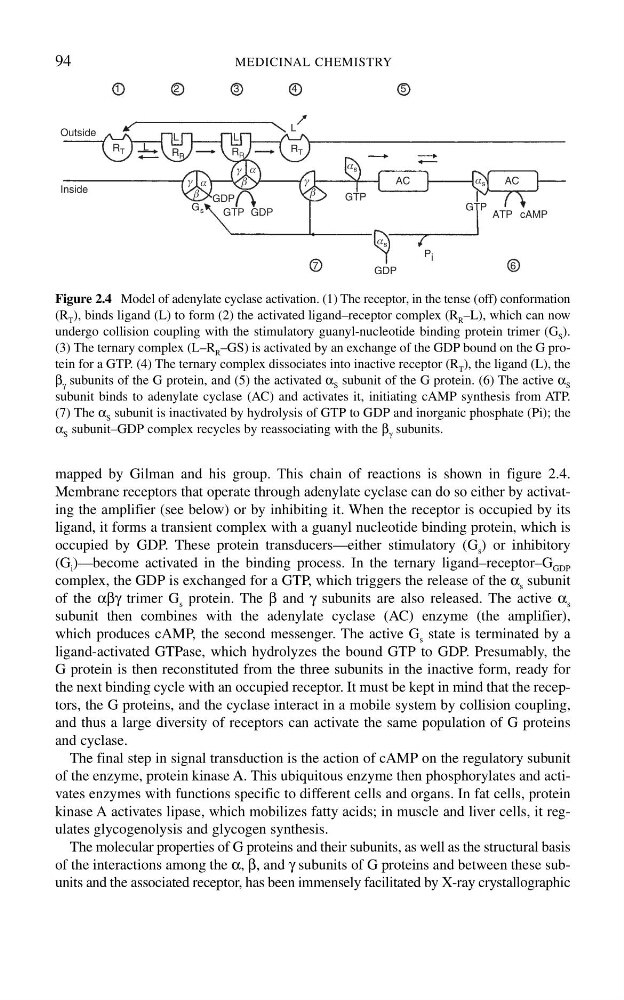
mapped by Gilman and his group. This chain of reactions is shown in figure 2.4.
Membrane receptors that operate through adenylate cyclase can do so either by activating the amplifier (see below) or by inhibiting it. When the receptor is occupied by its
ligand, it forms a transient complex with a guanyl nucleotide binding protein, which is
occupied by GDP. These protein transducers—either stimulatory (G s ) or inhibitory
(G i )—become activated in the binding process. In the ternary ligand–receptor–G GDP
complex, the GDP is exchanged for a GTP, which triggers the release of the α s subunit
of the αβγ trimer G s protein. The β and γ subunits are also released. The active α s
subunit then combines with the adenylate cyclase (AC) enzyme (the amplifier),
which produces cAMP, the second messenger. The active G s state is terminated by a
ligand-activated GTPase, which hydrolyzes the bound GTP to GDP. Presumably, the
G protein is then reconstituted from the three subunits in the inactive form, ready for
the next binding cycle with an occupied receptor. It must be kept in mind that the receptors, the G proteins, and the cyclase interact in a mobile system by collision coupling,
and thus a large diversity of receptors can activate the same population of G proteins
and cyclase.
The final step in signal transduction is the action of cAMP on the regulatory subunit
of the enzyme, protein kinase A. This ubiquitous enzyme then phosphorylates and activates enzymes with functions specific to different cells and organs. In fat cells, protein
kinase A activates lipase, which mobilizes fatty acids; in muscle and liver cells, it regulates glycogenolysis and glycogen synthesis.
The molecular properties of G proteins and their subunits, as well as the structural basis
of the interactions among the α, β, and γ subunits of G proteins and between these subunits and the associated receptor, has been immensely facilitated by X-ray crystallographic
94
MEDICINAL CHEMISTRY
1
2
3
4
7
6
5
Inside
Outside
GDP
GTP GDP
GTP
GTP
GDP
ATP cAMP
AC
AC
R T
R R
R R
R T
P i
a s
a s
a s
b
b
a
a
b
g
G s
L
L
L
g
g
L
Figure 2.4 Model of adenylate cyclase activation. (1) The receptor, in the tense (off) conformation
(R T ), binds ligand (L) to form (2) the activated ligand–receptor complex (R R –L), which can now
undergo collision coupling with the stimulatory guanyl-nucleotide binding protein trimer (G S ).
(3) The ternary complex (L–R R –GS) is activated by an exchange of the GDP bound on the G protein for a GTP. (4) The ternary complex dissociates into inactive receptor (R T ), the ligand (L), the
β γ subunits of the G protein, and (5) the activated α S subunit of the G protein. (6) The active α S
subunit binds to adenylate cyclase (AC) and activates it, initiating cAMP synthesis from ATP.
(7) The α S subunit is inactivated by hydrolysis of GTP to GDP and inorganic phosphate (Pi); the
α S subunit–GDP complex recycles by reassociating with the β γ subunits.
Membrane receptors that operate through adenylate cyclase can do so either by activating the amplifier (see below) or by inhibiting it. When the receptor is occupied by its
ligand, it forms a transient complex with a guanyl nucleotide binding protein, which is
occupied by GDP. These protein transducers—either stimulatory (G s ) or inhibitory
(G i )—become activated in the binding process. In the ternary ligand–receptor–G GDP
complex, the GDP is exchanged for a GTP, which triggers the release of the α s subunit
of the αβγ trimer G s protein. The β and γ subunits are also released. The active α s
subunit then combines with the adenylate cyclase (AC) enzyme (the amplifier),
which produces cAMP, the second messenger. The active G s state is terminated by a
ligand-activated GTPase, which hydrolyzes the bound GTP to GDP. Presumably, the
G protein is then reconstituted from the three subunits in the inactive form, ready for
the next binding cycle with an occupied receptor. It must be kept in mind that the receptors, the G proteins, and the cyclase interact in a mobile system by collision coupling,
and thus a large diversity of receptors can activate the same population of G proteins
and cyclase.
The final step in signal transduction is the action of cAMP on the regulatory subunit
of the enzyme, protein kinase A. This ubiquitous enzyme then phosphorylates and activates enzymes with functions specific to different cells and organs. In fat cells, protein
kinase A activates lipase, which mobilizes fatty acids; in muscle and liver cells, it regulates glycogenolysis and glycogen synthesis.
The molecular properties of G proteins and their subunits, as well as the structural basis
of the interactions among the α, β, and γ subunits of G proteins and between these subunits and the associated receptor, has been immensely facilitated by X-ray crystallographic
94
MEDICINAL CHEMISTRY
1
2
3
4
7
6
5
Inside
Outside
GDP
GTP GDP
GTP
GTP
GDP
ATP cAMP
AC
AC
R T
R R
R R
R T
P i
a s
a s
a s
b
b
a
a
b
g
G s
L
L
L
g
g
L
Figure 2.4 Model of adenylate cyclase activation. (1) The receptor, in the tense (off) conformation
(R T ), binds ligand (L) to form (2) the activated ligand–receptor complex (R R –L), which can now
undergo collision coupling with the stimulatory guanyl-nucleotide binding protein trimer (G S ).
(3) The ternary complex (L–R R –GS) is activated by an exchange of the GDP bound on the G protein for a GTP. (4) The ternary complex dissociates into inactive receptor (R T ), the ligand (L), the
β γ subunits of the G protein, and (5) the activated α S subunit of the G protein. (6) The active α S
subunit binds to adenylate cyclase (AC) and activates it, initiating cAMP synthesis from ATP.
(7) The α S subunit is inactivated by hydrolysis of GTP to GDP and inorganic phosphate (Pi); the
α S subunit–GDP complex recycles by reassociating with the β γ subunits.