

and miniaturization of devices. To date, the multi-disciplinary pressure ulcer round
team has been using ultrasonography for approximately 15 years. By analyzing the
vast amount of image data in detail, the evaluation viewpoint of pressure ulcer
ultrasound was determined, and it became possible to summarize the method of
evaluating pressure ulcers by ultrasound in the form of a flowchart (Fig. 7) (Aoi
et al., 2009). As can be seen from the normal image, the skin and subcutaneous tissue
are composed of layers, and the layer structure can be clearly confirmed by the
ultrasound images. If this layer structure is not clear, edema, granulation, necrotic
tissue, and so forth are presumed (Fig. 8). Moreover, if the tissue is replaced by scar
tissue after healing, the brightness may be increased as a whole. If the patient has a
pressure ulcer, the layer structure is often obscured by inflammatory edema. In
addition, cobblestone-like signs may be observed when edema findings increase.
The low-brightness area with an uneven internal structure is called a “cloud-like
image” and is likely to be necrotic tissue (Fig. 9).
The major difference between ultrasonography and other imaging devices, such
as CT and MRI, is that you can see only what you want to see. Ultrasonography
tends to cause artifacts (virtual images that do not actually exist) and requires the fine
adjustment of image quality, which is unsuitable for taking the entire image for
retrospective review. On the other hand, the great advantage of ultrasound is the
simplicity of the examination, the minimization of patient restraint, and the fact that
it can be evaluated in real-time. In addition, with the advancement of the device, it is
now much lighter and smaller than before, and the image quality has improved. It has
thus become possible to visualize a finer structure by using a high-frequency probe.
In this regard, it can be said that this is a novel nursing assessment technology
developed from the viewpoint of translational co-creation of information and knowledge between nursing science and engineering. In the future, we are developing
technologies to automatically acquire high-quality images and artificial intelligence
that automatically analyzes images to determine the degree of damage to deep
tissues.
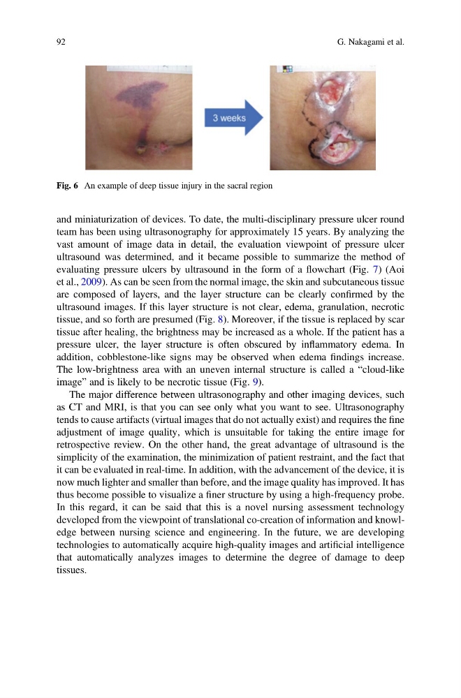
Fig. 6 An example of deep tissue injury in the sacral region
92
G. Nakagami et al.
team has been using ultrasonography for approximately 15 years. By analyzing the
vast amount of image data in detail, the evaluation viewpoint of pressure ulcer
ultrasound was determined, and it became possible to summarize the method of
evaluating pressure ulcers by ultrasound in the form of a flowchart (Fig. 7) (Aoi
et al., 2009). As can be seen from the normal image, the skin and subcutaneous tissue
are composed of layers, and the layer structure can be clearly confirmed by the
ultrasound images. If this layer structure is not clear, edema, granulation, necrotic
tissue, and so forth are presumed (Fig. 8). Moreover, if the tissue is replaced by scar
tissue after healing, the brightness may be increased as a whole. If the patient has a
pressure ulcer, the layer structure is often obscured by inflammatory edema. In
addition, cobblestone-like signs may be observed when edema findings increase.
The low-brightness area with an uneven internal structure is called a “cloud-like
image” and is likely to be necrotic tissue (Fig. 9).
The major difference between ultrasonography and other imaging devices, such
as CT and MRI, is that you can see only what you want to see. Ultrasonography
tends to cause artifacts (virtual images that do not actually exist) and requires the fine
adjustment of image quality, which is unsuitable for taking the entire image for
retrospective review. On the other hand, the great advantage of ultrasound is the
simplicity of the examination, the minimization of patient restraint, and the fact that
it can be evaluated in real-time. In addition, with the advancement of the device, it is
now much lighter and smaller than before, and the image quality has improved. It has
thus become possible to visualize a finer structure by using a high-frequency probe.
In this regard, it can be said that this is a novel nursing assessment technology
developed from the viewpoint of translational co-creation of information and knowledge between nursing science and engineering. In the future, we are developing
technologies to automatically acquire high-quality images and artificial intelligence
that automatically analyzes images to determine the degree of damage to deep
tissues.
Fig. 6 An example of deep tissue injury in the sacral region
92
G. Nakagami et al.