

is known as a proboscis hypha. The process of enzymatic secretion and cavity formation then is used to
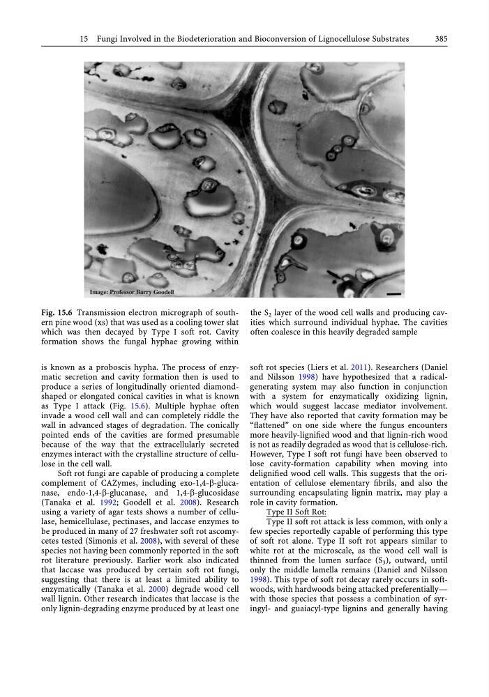
produce a series of longitudinally oriented diamondshaped or elongated conical cavities in what is known
as Type I attack (Fig. 15.6). Multiple hyphae often
invade a wood cell wall and can completely riddle the
wall in advanced stages of degradation. The conically
pointed ends of the cavities are formed presumable
because of the way that the extracellularly secreted
enzymes interact with the crystalline structure of cellulose in the cell wall.
Soft rot fungi are capable of producing a complete
complement of CAZymes, including exo-1,4-b-glucanase, endo-1,4-b-glucanase, and 1,4-b-glucosidase
(Tanaka et al. 1992; Goodell et al. 2008). Research
using a variety of agar tests shows a number of cellulase, hemicellulase, pectinases, and laccase enzymes to
be produced in many of 27 freshwater soft rot ascomycetes tested (Simonis et al. 2008), with several of these
species not having been commonly reported in the soft
rot literature previously. Earlier work also indicated
that laccase was produced by certain soft rot fungi,
suggesting that there is at least a limited ability to
enzymatically (Tanaka et al. 2000) degrade wood cell
wall lignin. Other research indicates that laccase is the
only lignin-degrading enzyme produced by at least one
soft rot species (Liers et al. 2011). Researchers (Daniel
and Nilsson 1998) have hypothesized that a radicalgenerating system may also function in conjunction
with a system for enzymatically oxidizing lignin,
which would suggest laccase mediator involvement.
They have also reported that cavity formation may be
“flattened” on one side where the fungus encounters
more heavily-lignified wood and that lignin-rich wood
is not as readily degraded as wood that is cellulose-rich.
However, Type I soft rot fungi have been observed to
lose cavity-formation capability when moving into
delignified wood cell walls. This suggests that the orientation of cellulose elementary fibrils, and also the
surrounding encapsulating lignin matrix, may play a
role in cavity formation.
Type II Soft Rot:
Type II soft rot attack is less common, with only a
few species reportedly capable of performing this type
of soft rot alone. Type II soft rot appears similar to
white rot at the microscale, as the wood cell wall is
thinned from the lumen surface (S 3 ), outward, until
only the middle lamella remains (Daniel and Nilsson
1998). This type of soft rot decay rarely occurs in softwoods, with hardwoods being attacked preferentially—
with those species that possess a combination of syringyl- and guaiacyl-type lignins and generally having
Fig. 15.6 Transmission electron micrograph of southern pine wood (xs) that was used as a cooling tower slat
which was then decayed by Type I soft rot. Cavity
formation shows the fungal hyphae growing within
the S 2 layer of the wood cell walls and producing cavities which surround individual hyphae. The cavities
often coalesce in this heavily degraded sample
15 Fungi Involved in the Biodeterioration and Bioconversion of Lignocellulose Substrates
385
produce a series of longitudinally oriented diamondshaped or elongated conical cavities in what is known
as Type I attack (Fig. 15.6). Multiple hyphae often
invade a wood cell wall and can completely riddle the
wall in advanced stages of degradation. The conically
pointed ends of the cavities are formed presumable
because of the way that the extracellularly secreted
enzymes interact with the crystalline structure of cellulose in the cell wall.
Soft rot fungi are capable of producing a complete
complement of CAZymes, including exo-1,4-b-glucanase, endo-1,4-b-glucanase, and 1,4-b-glucosidase
(Tanaka et al. 1992; Goodell et al. 2008). Research
using a variety of agar tests shows a number of cellulase, hemicellulase, pectinases, and laccase enzymes to
be produced in many of 27 freshwater soft rot ascomycetes tested (Simonis et al. 2008), with several of these
species not having been commonly reported in the soft
rot literature previously. Earlier work also indicated
that laccase was produced by certain soft rot fungi,
suggesting that there is at least a limited ability to
enzymatically (Tanaka et al. 2000) degrade wood cell
wall lignin. Other research indicates that laccase is the
only lignin-degrading enzyme produced by at least one
soft rot species (Liers et al. 2011). Researchers (Daniel
and Nilsson 1998) have hypothesized that a radicalgenerating system may also function in conjunction
with a system for enzymatically oxidizing lignin,
which would suggest laccase mediator involvement.
They have also reported that cavity formation may be
“flattened” on one side where the fungus encounters
more heavily-lignified wood and that lignin-rich wood
is not as readily degraded as wood that is cellulose-rich.
However, Type I soft rot fungi have been observed to
lose cavity-formation capability when moving into
delignified wood cell walls. This suggests that the orientation of cellulose elementary fibrils, and also the
surrounding encapsulating lignin matrix, may play a
role in cavity formation.
Type II Soft Rot:
Type II soft rot attack is less common, with only a
few species reportedly capable of performing this type
of soft rot alone. Type II soft rot appears similar to
white rot at the microscale, as the wood cell wall is
thinned from the lumen surface (S 3 ), outward, until
only the middle lamella remains (Daniel and Nilsson
1998). This type of soft rot decay rarely occurs in softwoods, with hardwoods being attacked preferentially—
with those species that possess a combination of syringyl- and guaiacyl-type lignins and generally having
Fig. 15.6 Transmission electron micrograph of southern pine wood (xs) that was used as a cooling tower slat
which was then decayed by Type I soft rot. Cavity
formation shows the fungal hyphae growing within
the S 2 layer of the wood cell walls and producing cavities which surround individual hyphae. The cavities
often coalesce in this heavily degraded sample
15 Fungi Involved in the Biodeterioration and Bioconversion of Lignocellulose Substrates
385