

PhyB-PIF3 (PIF6)
PhyA-PIF3
VVD-VVD (LightOn)
WC1-VVD (FUN-LOV)
CRY2-CIB1
PhyA-FHY1
BphP1-PpsR2
UVR8-COP1
FKF1-GI
EL222-EL222
Photoreceptor (PhR) -
Interacting Protien (IP)
Different PhR-PhR or
PhR dimerization
No interaction
DBD
AD
DBD
DBD
AD
AD
AD
PhR
PhR
PhR
IP
PhR
PhR
IP
i
o
g
i
o
g
i
o
g
Light
Dark
B
A
C
goi
IP
dCas9
AD
PhR
goi
IP
dCas9
AD
PhR
Interaction
Light
Dark
AsLOV2 or AtLOV2
D
PhR
poi
IP
Light
Dark
poi
IP
PM
PM
PhR
PM
PhR
p o i
P h R
P h R
or
or
p o i
Light
Dark
AsLOV2 or AtLOV2
J -helix
E
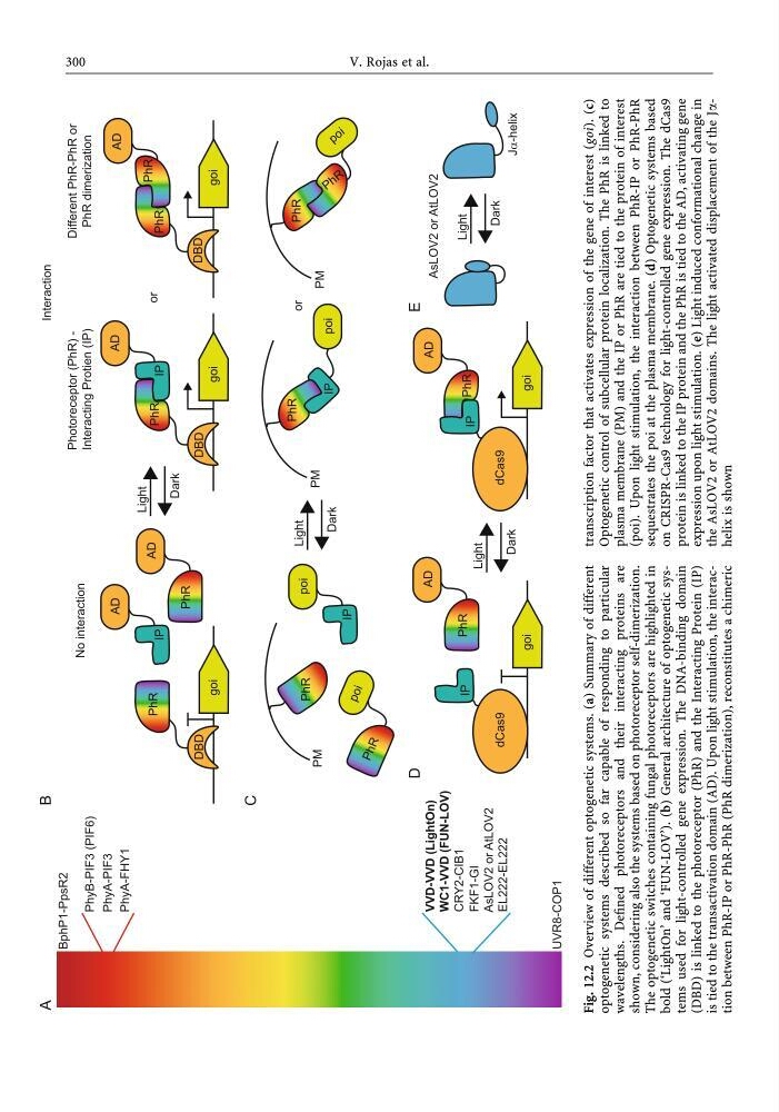
Fig. 12.2 Overview of different optogenetic systems. (a) Summary of different
optogenetic systems described so far capable of responding to particular
wavelengths. Defined photoreceptors and their interacting proteins are
shown, considering also the systems based on photoreceptor self-dimerization.
The optogenetic switches containing fungal photoreceptors are highlighted in
bold (‘LightOn’ and ‘FUN-LOV’). (b) General architecture of optogenetic systems used for light-controlled gene expression. The DNA-binding domain
(DBD) is linked to the photoreceptor (PhR) and the Interacting Protein (IP)
is tied to the transactivation domain (AD). Upon light stimulation, the interaction between PhR-IP or PhR-PhR (PhR dimerization), reconstitutes a chimeric
transcription factor that activates expression of the gene of interest (goi). (c)
Optogenetic control of subcellular protein localization. The PhR is linked to
plasma membrane (PM) and the IP or PhR are tied to the protein of interest
(poi). Upon light stimulation, the interaction between PhR-IP or PhR-PhR
sequestrates the poi at the plasma membrane. (d) Optogenetic systems based
on CRISPR-Cas9 technology for light-controlled gene expression. The dCas9
protein is linked to the IP protein and the PhR is tied to the AD, activating gene
expression upon light stimulation. (e) Light induced conformational change in
the AsLOV2 or AtLOV2 domains. The light activated displacement of the Jahelix is shown
300
V. Rojas et al.
PhyA-PIF3
VVD-VVD (LightOn)
WC1-VVD (FUN-LOV)
CRY2-CIB1
PhyA-FHY1
BphP1-PpsR2
UVR8-COP1
FKF1-GI
EL222-EL222
Photoreceptor (PhR) -
Interacting Protien (IP)
Different PhR-PhR or
PhR dimerization
No interaction
DBD
AD
DBD
DBD
AD
AD
AD
PhR
PhR
PhR
IP
PhR
PhR
IP
i
o
g
i
o
g
i
o
g
Light
Dark
B
A
C
goi
IP
dCas9
AD
PhR
goi
IP
dCas9
AD
PhR
Interaction
Light
Dark
AsLOV2 or AtLOV2
D
PhR
poi
IP
Light
Dark
poi
IP
PM
PM
PhR
PM
PhR
p o i
P h R
P h R
or
or
p o i
Light
Dark
AsLOV2 or AtLOV2
J -helix
E
Fig. 12.2 Overview of different optogenetic systems. (a) Summary of different
optogenetic systems described so far capable of responding to particular
wavelengths. Defined photoreceptors and their interacting proteins are
shown, considering also the systems based on photoreceptor self-dimerization.
The optogenetic switches containing fungal photoreceptors are highlighted in
bold (‘LightOn’ and ‘FUN-LOV’). (b) General architecture of optogenetic systems used for light-controlled gene expression. The DNA-binding domain
(DBD) is linked to the photoreceptor (PhR) and the Interacting Protein (IP)
is tied to the transactivation domain (AD). Upon light stimulation, the interaction between PhR-IP or PhR-PhR (PhR dimerization), reconstitutes a chimeric
transcription factor that activates expression of the gene of interest (goi). (c)
Optogenetic control of subcellular protein localization. The PhR is linked to
plasma membrane (PM) and the IP or PhR are tied to the protein of interest
(poi). Upon light stimulation, the interaction between PhR-IP or PhR-PhR
sequestrates the poi at the plasma membrane. (d) Optogenetic systems based
on CRISPR-Cas9 technology for light-controlled gene expression. The dCas9
protein is linked to the IP protein and the PhR is tied to the AD, activating gene
expression upon light stimulation. (e) Light induced conformational change in
the AsLOV2 or AtLOV2 domains. The light activated displacement of the Jahelix is shown
300
V. Rojas et al.