

not possess fatty acid synthetases (FAS; Wewer
et al. 2014), resulting to be auxotroph for this
essential group of compounds which are totally
received from the host plant (Luginbuehl et al.
2017; Jiang et al. 2017; Keymer et al. 2017).
Interestingly, Tisserant et al. (2013) firstly
pointed to the almost total lack of genes encoding plant cell wall-degrading enzymes as well
as of genes involved in toxin and thiamine
synthesis. The limited number of cell walldegrading enzymes is currently considered
one of the signatures of AM fungal genomes,
since this feature has been found in all other
genomes sequenced later: R. clarus (Kobayashi
et al. 2018), Rhizophagus cerebriforme, and Rhizophagus diaphanum (Morin et al. 2018) all
belonging to the same Rhizophagus genus, as
well as members of the Diversisporales, Diversispora epigea (Sun et al. 2018), Gigaspora rosea
(Morin et al. 2018), and Gigaspora margarita
(Venice et al. 2019). Taken in the whole, these
results highlight a still unsolved question: how
do AM fungi penetrate within their host tissue,
being for a large part of their life cycle intracellular endophytes? How do they cross the plant
cell wall? On one hand, the limited number of
such hydrolytic enzymes well fits with the life
strategy of AM fungi: they do not activate plant
defenses, since they have to keep their host
alive and prone to accommodate the fungus,
but, on the other hand, they enter the root
cells probably thanks to a finely tuned regulation of the plant metabolism. Among the battery of mycorrhiza-induced secreted proteins
which are mainly expressed in symbiotic tissues (Tisserant et al. 2013; Se ˛dzielewska Toro
and Brachmann 2016), some of them can act as
effectors which might manipulate plant regulatory pathways (see next paragraph). It can be
hypothesized that some effectors may induce
the weakening and swelling of plant cell walls,
which may compensate for the absence of
hydrolytic enzymes and is indeed observed by
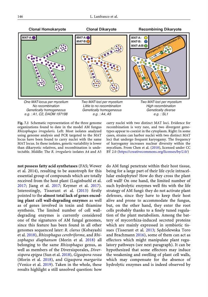
Fig. 7.1 Schematic representation of the three genome
organizations found to date in the model AM fungus
Rhizophagus irregularis. Left: Most isolates analyzed
using genome analysis and PCR targeted to the MAT
locus have been found to carry nuclei with the same
MAT locus. In these isolates, genetic variability is lower
than dikaryotic relatives, and recombination is undetectable. Middle: The R. irregularis isolates A4 and A5
carry nuclei with two distinct MAT loci. Evidence for
recombination is very rare, and two divergent genotypes appear to coexist in the cytoplasm. Right: In some
cases, strains can harbor nuclei with two distinct MAT
loci that undergo frequent karyogamy. The frequency
of karyogamy increases nuclear diversity within the
mycelium. From Chen et al. (2018), licensed under CC
BY 2.0 (https://creativecommons.org/licenses/by/2.0/)
146
L. Lanfranco et al.
et al. 2014), resulting to be auxotroph for this
essential group of compounds which are totally
received from the host plant (Luginbuehl et al.
2017; Jiang et al. 2017; Keymer et al. 2017).
Interestingly, Tisserant et al. (2013) firstly
pointed to the almost total lack of genes encoding plant cell wall-degrading enzymes as well
as of genes involved in toxin and thiamine
synthesis. The limited number of cell walldegrading enzymes is currently considered
one of the signatures of AM fungal genomes,
since this feature has been found in all other
genomes sequenced later: R. clarus (Kobayashi
et al. 2018), Rhizophagus cerebriforme, and Rhizophagus diaphanum (Morin et al. 2018) all
belonging to the same Rhizophagus genus, as
well as members of the Diversisporales, Diversispora epigea (Sun et al. 2018), Gigaspora rosea
(Morin et al. 2018), and Gigaspora margarita
(Venice et al. 2019). Taken in the whole, these
results highlight a still unsolved question: how
do AM fungi penetrate within their host tissue,
being for a large part of their life cycle intracellular endophytes? How do they cross the plant
cell wall? On one hand, the limited number of
such hydrolytic enzymes well fits with the life
strategy of AM fungi: they do not activate plant
defenses, since they have to keep their host
alive and prone to accommodate the fungus,
but, on the other hand, they enter the root
cells probably thanks to a finely tuned regulation of the plant metabolism. Among the battery of mycorrhiza-induced secreted proteins
which are mainly expressed in symbiotic tissues (Tisserant et al. 2013; Se ˛dzielewska Toro
and Brachmann 2016), some of them can act as
effectors which might manipulate plant regulatory pathways (see next paragraph). It can be
hypothesized that some effectors may induce
the weakening and swelling of plant cell walls,
which may compensate for the absence of
hydrolytic enzymes and is indeed observed by
Fig. 7.1 Schematic representation of the three genome
organizations found to date in the model AM fungus
Rhizophagus irregularis. Left: Most isolates analyzed
using genome analysis and PCR targeted to the MAT
locus have been found to carry nuclei with the same
MAT locus. In these isolates, genetic variability is lower
than dikaryotic relatives, and recombination is undetectable. Middle: The R. irregularis isolates A4 and A5
carry nuclei with two distinct MAT loci. Evidence for
recombination is very rare, and two divergent genotypes appear to coexist in the cytoplasm. Right: In some
cases, strains can harbor nuclei with two distinct MAT
loci that undergo frequent karyogamy. The frequency
of karyogamy increases nuclear diversity within the
mycelium. From Chen et al. (2018), licensed under CC
BY 2.0 (https://creativecommons.org/licenses/by/2.0/)
146
L. Lanfranco et al.