

discrete phylogenetic subtypes which include
the R0 motifs of the corresponding NLR-like
proteins, suggesting that a signal-transducing
specificity exists for each subtype. Five different
HRAM subtypes have been described, which
share a pattern of alternating hydrophobic/
polar (charged) residues (Daskalov et al.
2015a). The common primary sequence pattern
and conservation of functionally important
residues for the b-solenoid fold of HET-S suggest that the HRAMs adopt an overall similar
amyloid fold. The discovery of the HRAMs
establishes a superfamily of signal-transducing
amyloids, which has prompted comparisons
with the death domains superfamily (CARD,
PYD, DD, and DED), involved in the control
of apoptosis and inflammatory cell death in
metazoans (Park et al. 2007). The death-fold
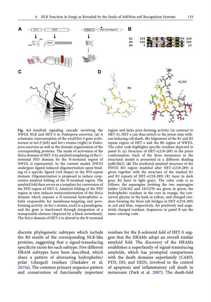
Fig. 6.3 Amyloid signaling cascade involving the
NWD2 NLR and HET-S in Podospora anserina. (a) A
schematic representation of the nwd2/het-S gene architecture in het-S (left) and het-s strains (right) in Podospora anserina as well as the domain organization of the
corresponding proteins. The mode of activation of the
HeLo domain of HET-S by amyloid templating of the Cterminal PFD domain by the N-terminal region of
NWD2 is represented. In the current model, NWD2
undergoes ligand-induced oligomerization upon binding of a specific ligand (red shape) in the WD-repeat
domain. Oligomerization is proposed to induce cooperative amyloid folding of the N-terminal region. The
amyloid fold then serves as a template for conversion of
the PFD region of HET-S. Amyloid folding of the PFD
region in turn induces transconformation of the HeLo
domain which exposes a N-terminal hydrophobic ahelix responsible for membrane-targeting and poreforming activity. In het-s strains, nwd2 is a pseudogene,
and the gene is inactivated through integration of a
transposable element (depicted by a black arrowhead).
The HeLo domain of HET-s in altered in the N-terminal
region and lacks pore-forming activity (in contrast to
HET-S), HET-s can thus switch to the prion state without inducing cell death. (b) Alignment of the R1 and R2
repeat region of HET-s and the R0 region of NWD2.
The color code highlights specific residues depicted in
panel D. (c) Structure of HET-s(218-289) in the prion
conformation. Each of the three monomers in the
structural model is presented in a different shading
(pdb:2kj3). (d) The predicted amyloid structure of the
NWD2 RO region modeled after HET-s(218-289) is
given together with the structure of the stacked R1
and R2 repeats of HET-s(218-289) (R1 layer in dark
gray, R2 layer in light gray). The color code is as
follows: the asparagine forming the two asparagine
ladder (226/262 and 243/279) are given in green, the
hydrophobic residues in the core in orange, the conserved glycine in the kink in yellow, and charged residues forming the three salt bridges in HET-s(218-289)
in red and blue, respectively, for positively and negatively charged residues. Sequences in panel B use the
same coloring code
6 NLR Function in Fungi as Revealed by the Study of Self/Non-self Recognition Systems
133
the R0 motifs of the corresponding NLR-like
proteins, suggesting that a signal-transducing
specificity exists for each subtype. Five different
HRAM subtypes have been described, which
share a pattern of alternating hydrophobic/
polar (charged) residues (Daskalov et al.
2015a). The common primary sequence pattern
and conservation of functionally important
residues for the b-solenoid fold of HET-S suggest that the HRAMs adopt an overall similar
amyloid fold. The discovery of the HRAMs
establishes a superfamily of signal-transducing
amyloids, which has prompted comparisons
with the death domains superfamily (CARD,
PYD, DD, and DED), involved in the control
of apoptosis and inflammatory cell death in
metazoans (Park et al. 2007). The death-fold
Fig. 6.3 Amyloid signaling cascade involving the
NWD2 NLR and HET-S in Podospora anserina. (a) A
schematic representation of the nwd2/het-S gene architecture in het-S (left) and het-s strains (right) in Podospora anserina as well as the domain organization of the
corresponding proteins. The mode of activation of the
HeLo domain of HET-S by amyloid templating of the Cterminal PFD domain by the N-terminal region of
NWD2 is represented. In the current model, NWD2
undergoes ligand-induced oligomerization upon binding of a specific ligand (red shape) in the WD-repeat
domain. Oligomerization is proposed to induce cooperative amyloid folding of the N-terminal region. The
amyloid fold then serves as a template for conversion of
the PFD region of HET-S. Amyloid folding of the PFD
region in turn induces transconformation of the HeLo
domain which exposes a N-terminal hydrophobic ahelix responsible for membrane-targeting and poreforming activity. In het-s strains, nwd2 is a pseudogene,
and the gene is inactivated through integration of a
transposable element (depicted by a black arrowhead).
The HeLo domain of HET-s in altered in the N-terminal
region and lacks pore-forming activity (in contrast to
HET-S), HET-s can thus switch to the prion state without inducing cell death. (b) Alignment of the R1 and R2
repeat region of HET-s and the R0 region of NWD2.
The color code highlights specific residues depicted in
panel D. (c) Structure of HET-s(218-289) in the prion
conformation. Each of the three monomers in the
structural model is presented in a different shading
(pdb:2kj3). (d) The predicted amyloid structure of the
NWD2 RO region modeled after HET-s(218-289) is
given together with the structure of the stacked R1
and R2 repeats of HET-s(218-289) (R1 layer in dark
gray, R2 layer in light gray). The color code is as
follows: the asparagine forming the two asparagine
ladder (226/262 and 243/279) are given in green, the
hydrophobic residues in the core in orange, the conserved glycine in the kink in yellow, and charged residues forming the three salt bridges in HET-s(218-289)
in red and blue, respectively, for positively and negatively charged residues. Sequences in panel B use the
same coloring code
6 NLR Function in Fungi as Revealed by the Study of Self/Non-self Recognition Systems
133