

506
T. Borowski and M. Szaleniec
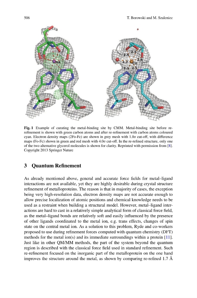
Fig. 1 Example of curating the metal-binding site by CMM. Metal-binding site before rerefinement is shown with green carbon atoms and after re-refinement with carbon atoms coloured
cyan. Electron density maps (2Fo-Fc) are shown in grey mesh with 1.0σ cut-off, with difference
maps (Fo-Fc) shown in green and red mesh with 4.0σ cut-off. In the re-refined structure, only one
of the two alternative glycerol molecules is shown for clarity. Reprinted with permission from [8].
Copyright 2013 Springer Nature
3 Quantum Refinement
As already mentioned above, general and accurate force fields for metal–ligand
interactions are not available, yet they are highly desirable during crystal structure
refinement of metalloproteins. The reason is that in majority of cases, the exception
being very high-resolution data, electron density maps are not accurate enough to
allow precise localization of atomic positions and chemical knowledge needs to be
used as a restraint when building a structural model. However, metal–ligand interactions are hard to cast in a relatively simple analytical form of classical force field,
as the metal–ligand bonds are relatively soft and easily influenced by the presence
of other ligands coordinated to the metal ion, e.g. trans effects, changes of spin
state on the central metal ion. As a solution to this problem, Ryde and co-workers
proposed to use during refinement forces computed with quantum chemistry (DFT)
methods for the metal ion(s) and its immediate surroundings within a protein [11].
Just like in other QM/MM methods, the part of the system beyond the quantum
region is described with the classical force field used in standard refinement. Such
re-refinement focused on the inorganic part of the metalloprotein on the one hand
improves the structure around the metal, as shown by comparing re-refined 1.7 Å
T. Borowski and M. Szaleniec
Fig. 1 Example of curating the metal-binding site by CMM. Metal-binding site before rerefinement is shown with green carbon atoms and after re-refinement with carbon atoms coloured
cyan. Electron density maps (2Fo-Fc) are shown in grey mesh with 1.0σ cut-off, with difference
maps (Fo-Fc) shown in green and red mesh with 4.0σ cut-off. In the re-refined structure, only one
of the two alternative glycerol molecules is shown for clarity. Reprinted with permission from [8].
Copyright 2013 Springer Nature
3 Quantum Refinement
As already mentioned above, general and accurate force fields for metal–ligand
interactions are not available, yet they are highly desirable during crystal structure
refinement of metalloproteins. The reason is that in majority of cases, the exception
being very high-resolution data, electron density maps are not accurate enough to
allow precise localization of atomic positions and chemical knowledge needs to be
used as a restraint when building a structural model. However, metal–ligand interactions are hard to cast in a relatively simple analytical form of classical force field,
as the metal–ligand bonds are relatively soft and easily influenced by the presence
of other ligands coordinated to the metal ion, e.g. trans effects, changes of spin
state on the central metal ion. As a solution to this problem, Ryde and co-workers
proposed to use during refinement forces computed with quantum chemistry (DFT)
methods for the metal ion(s) and its immediate surroundings within a protein [11].
Just like in other QM/MM methods, the part of the system beyond the quantum
region is described with the classical force field used in standard refinement. Such
re-refinement focused on the inorganic part of the metalloprotein on the one hand
improves the structure around the metal, as shown by comparing re-refined 1.7 Å