

505
15 (How to) Profit from Molecular Dynamics-based Ensemble Docking
mutation I223R (PDB codes 3NSS, 3TI5 and 4B7M [28–30]). The starting structure of the mutant I223R has an alternative backbone conformation at a loop region
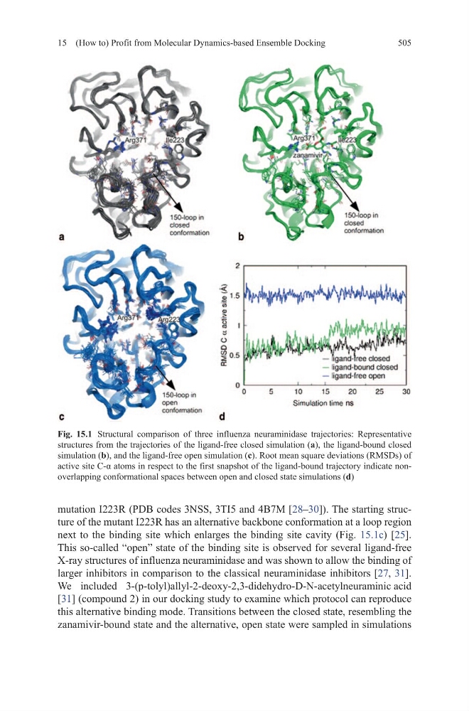
next to the binding site which enlarges the binding site cavity (Fig. 15.1c) [25].
This so-called “open” state of the binding site is observed for several ligand-free
X-ray structures of influenza neuraminidase and was shown to allow the binding of
larger inhibitors in comparison to the classical neuraminidase inhibitors [27, 31].
We included 3-(p-tolyl)allyl-2-deoxy-2,3-didehydro-D-N-acetylneuraminic acid
[31] (compound 2) in our docking study to examine which protocol can reproduce
this alternative binding mode. Transitions between the closed state, resembling the
zanamivir-bound state and the alternative, open state were sampled in simulations
Fig. 15.1 Structural comparison of three influenza neuraminidase trajectories: Representative
structures from the trajectories of the ligand-free closed simulation (a), the ligand-bound closed
simulation (b), and the ligand-free open simulation (c). Root mean square deviations (RMSDs) of
active site C-α atoms in respect to the first snapshot of the ligand-bound trajectory indicate nonoverlapping conformational spaces between open and closed state simulations (d)
15 (How to) Profit from Molecular Dynamics-based Ensemble Docking
mutation I223R (PDB codes 3NSS, 3TI5 and 4B7M [28–30]). The starting structure of the mutant I223R has an alternative backbone conformation at a loop region
next to the binding site which enlarges the binding site cavity (Fig. 15.1c) [25].
This so-called “open” state of the binding site is observed for several ligand-free
X-ray structures of influenza neuraminidase and was shown to allow the binding of
larger inhibitors in comparison to the classical neuraminidase inhibitors [27, 31].
We included 3-(p-tolyl)allyl-2-deoxy-2,3-didehydro-D-N-acetylneuraminic acid
[31] (compound 2) in our docking study to examine which protocol can reproduce
this alternative binding mode. Transitions between the closed state, resembling the
zanamivir-bound state and the alternative, open state were sampled in simulations
Fig. 15.1 Structural comparison of three influenza neuraminidase trajectories: Representative
structures from the trajectories of the ligand-free closed simulation (a), the ligand-bound closed
simulation (b), and the ligand-free open simulation (c). Root mean square deviations (RMSDs) of
active site C-α atoms in respect to the first snapshot of the ligand-bound trajectory indicate nonoverlapping conformational spaces between open and closed state simulations (d)