

123
The molecular docking study was performed with the Glide program. One
should mentioned that ADV is a prodrug, and due to its bioactive form, ADV diphosphate (ADV-DP), which is generated through stepwise phosphorylation after
the oral administration of ADV, was modeled,. The 3D conformation of ADV-DP
was constructed according to the coordinates of the ligand, TDV-DP, which was
deposited in the template structure, by deleting a methyl group.
The molecular docking was performed using standard precision protocols with
default parameters. The docking poses were ranked by their glide scores, and the
best predicted conformation in each system was used for the subsequent molecular
dynamics simulation and binding energy analysis.
Classical molecular dynamics simulations of docked conformation-HBV polymerase complexes were performed using the AMBER 9.0 suite [148]. The electrostatic potential of ADV-DP was computed with Gaussian03 at the HF/6-31G*
level [149], then the charges were assigned using the RESP (restrained electrostatic
potential) methodology [150]. The charges and force field parameters for ADV-DP
were generated by an Antechamber [151]. The polyphosphate parameters of ADVDP in studied system were identical to those developed by Meagher et al. [152].
The AMBER03 force field [142] and the general AMBER force field (GAFF) [153]
were chosen to create the potential of the proteins and ADV-DP, respectively. The
systems were then solvated in a truncated octahedral box of TIP3P water molecules
10 Å away from the protein. Counter-ions of Cl
−
were then added to obtain the electrostatic neutrality of the systems. The ADV-DP binding free energies (ΔG bind ) HBV
pols of B and C genotypes were calculated using the MM-PBSA method [154]. The
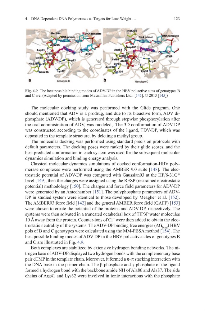
best possible binding modes of ADV-DP in the HBV pol active sites of genotypes B
and C are illustrated in Fig. 4.9.
Both complexes are stabilized by extensive hydrogen bonding networks. The nitrogen base of ADV-DP displayed two hydrogen bonds with the complementary base
pair dTMP in the template chain. Moreover, it formed a π–π stacking interaction with
the DNA base in the primer chain. The β-phosphate and γ-phosphate of the ligand
formed a hydrogen bond with the backbone amide NH of Ala86 and Ala87. The side
chains of Arg41 and Lys32 were involved in ionic interactions with the phosphate
4 DNA Dependent DNA Polymerases as Targets for Low-Weight …
Fig. 4.9 The best possible binding modes of ADV-DP in the HBV pol active sites of genotypes B
and C are. (Adapted by permission from Macmillan Publishers Ltd.: [145]. © 2013 [145])
The molecular docking study was performed with the Glide program. One
should mentioned that ADV is a prodrug, and due to its bioactive form, ADV diphosphate (ADV-DP), which is generated through stepwise phosphorylation after
the oral administration of ADV, was modeled,. The 3D conformation of ADV-DP
was constructed according to the coordinates of the ligand, TDV-DP, which was
deposited in the template structure, by deleting a methyl group.
The molecular docking was performed using standard precision protocols with
default parameters. The docking poses were ranked by their glide scores, and the
best predicted conformation in each system was used for the subsequent molecular
dynamics simulation and binding energy analysis.
Classical molecular dynamics simulations of docked conformation-HBV polymerase complexes were performed using the AMBER 9.0 suite [148]. The electrostatic potential of ADV-DP was computed with Gaussian03 at the HF/6-31G*
level [149], then the charges were assigned using the RESP (restrained electrostatic
potential) methodology [150]. The charges and force field parameters for ADV-DP
were generated by an Antechamber [151]. The polyphosphate parameters of ADVDP in studied system were identical to those developed by Meagher et al. [152].
The AMBER03 force field [142] and the general AMBER force field (GAFF) [153]
were chosen to create the potential of the proteins and ADV-DP, respectively. The
systems were then solvated in a truncated octahedral box of TIP3P water molecules
10 Å away from the protein. Counter-ions of Cl
−
were then added to obtain the electrostatic neutrality of the systems. The ADV-DP binding free energies (ΔG bind ) HBV
pols of B and C genotypes were calculated using the MM-PBSA method [154]. The
best possible binding modes of ADV-DP in the HBV pol active sites of genotypes B
and C are illustrated in Fig. 4.9.
Both complexes are stabilized by extensive hydrogen bonding networks. The nitrogen base of ADV-DP displayed two hydrogen bonds with the complementary base
pair dTMP in the template chain. Moreover, it formed a π–π stacking interaction with
the DNA base in the primer chain. The β-phosphate and γ-phosphate of the ligand
formed a hydrogen bond with the backbone amide NH of Ala86 and Ala87. The side
chains of Arg41 and Lys32 were involved in ionic interactions with the phosphate
4 DNA Dependent DNA Polymerases as Targets for Low-Weight …
Fig. 4.9 The best possible binding modes of ADV-DP in the HBV pol active sites of genotypes B
and C are. (Adapted by permission from Macmillan Publishers Ltd.: [145]. © 2013 [145])