

control [7, 8]. Further, this process is only applicable to cells in
suspension.
Localized electroporation is an alternative to bulk electroporation utilizing adherent cells on a perforated surface. The perforated
surface allows the cells to adhere and grow naturally, thus improving the health of the cells and providing a better reference to cells as
they adhere in the body [9]. An electric field is generated between
electrodes on each side of the perforated surface with electrical
conduction only occurring through nanochannels. Cells will be
porated only in the membrane areas exposed to the electrical field
through the pores on the perforated surface. The reduction in
surface area of the membrane exposed to the electrical field means
lower voltages can be used, thus smaller and more uniform electric
fields can be generated, and smaller pores are produced in the cell
membrane, all resulting in increased viability and transfection efficiency [10, 11]. Moreover, the adherent state of the cells allows
them to be repeatedly transfected with varying molecules and
observed over a prolonged period.
This chapter discusses the creation and use of a simple and
cost-effective lab-on-a-chip microdevice for localized electroporation. Utilizing a porous membrane with nanopores (size ranging
from 50 nm to 200 nm), the device applies voltage to a small
patch of the cell membrane to effectively reduce the exposed area
and thus the amount of voltage required to induce pore formation
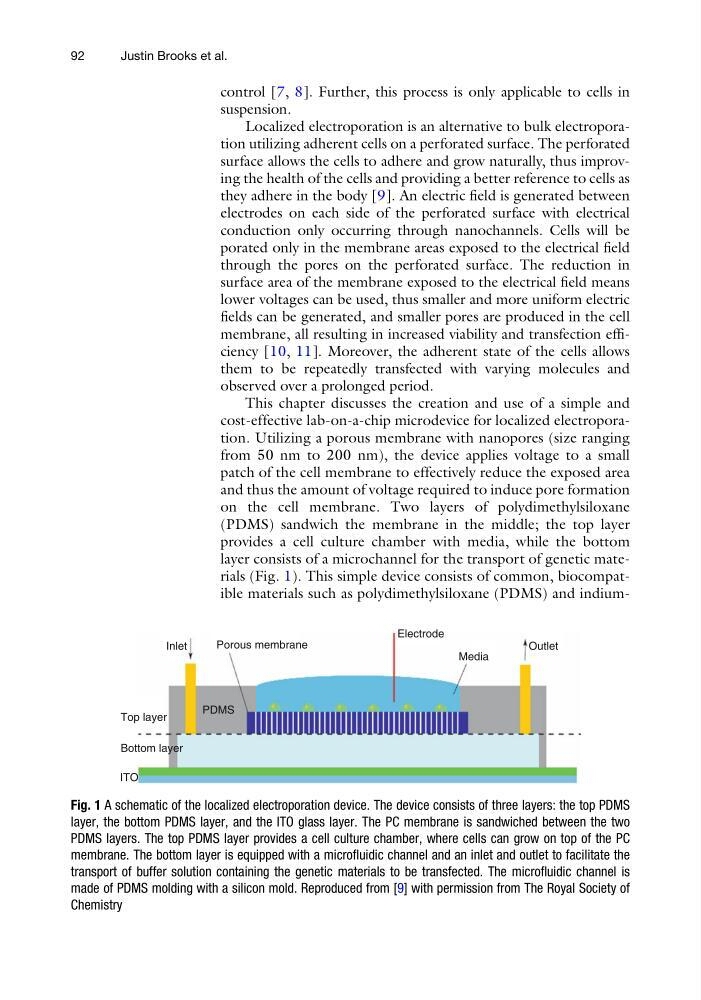
on the cell membrane. Two layers of polydimethylsiloxane
(PDMS) sandwich the membrane in the middle; the top layer
provides a cell culture chamber with media, while the bottom
layer consists of a microchannel for the transport of genetic materials (Fig. 1). This simple device consists of common, biocompatible materials such as polydimethylsiloxane (PDMS) and indiumITO
Bottom layer
Top layer
PDMS
Inlet
Porous membrane
Electrode
Media
Outlet
Fig. 1 A schematic of the localized electroporation device. The device consists of three layers: the top PDMS
layer, the bottom PDMS layer, and the ITO glass layer. The PC membrane is sandwiched between the two
PDMS layers. The top PDMS layer provides a cell culture chamber, where cells can grow on top of the PC
membrane. The bottom layer is equipped with a microfluidic channel and an inlet and outlet to facilitate the
transport of buffer solution containing the genetic materials to be transfected. The microfluidic channel is
made of PDMS molding with a silicon mold. Reproduced from [9] with permission from The Royal Society of
Chemistry
92
Justin Brooks et al.
suspension.
Localized electroporation is an alternative to bulk electroporation utilizing adherent cells on a perforated surface. The perforated
surface allows the cells to adhere and grow naturally, thus improving the health of the cells and providing a better reference to cells as
they adhere in the body [9]. An electric field is generated between
electrodes on each side of the perforated surface with electrical
conduction only occurring through nanochannels. Cells will be
porated only in the membrane areas exposed to the electrical field
through the pores on the perforated surface. The reduction in
surface area of the membrane exposed to the electrical field means
lower voltages can be used, thus smaller and more uniform electric
fields can be generated, and smaller pores are produced in the cell
membrane, all resulting in increased viability and transfection efficiency [10, 11]. Moreover, the adherent state of the cells allows
them to be repeatedly transfected with varying molecules and
observed over a prolonged period.
This chapter discusses the creation and use of a simple and
cost-effective lab-on-a-chip microdevice for localized electroporation. Utilizing a porous membrane with nanopores (size ranging
from 50 nm to 200 nm), the device applies voltage to a small
patch of the cell membrane to effectively reduce the exposed area
and thus the amount of voltage required to induce pore formation
on the cell membrane. Two layers of polydimethylsiloxane
(PDMS) sandwich the membrane in the middle; the top layer
provides a cell culture chamber with media, while the bottom
layer consists of a microchannel for the transport of genetic materials (Fig. 1). This simple device consists of common, biocompatible materials such as polydimethylsiloxane (PDMS) and indiumITO
Bottom layer
Top layer
PDMS
Inlet
Porous membrane
Electrode
Media
Outlet
Fig. 1 A schematic of the localized electroporation device. The device consists of three layers: the top PDMS
layer, the bottom PDMS layer, and the ITO glass layer. The PC membrane is sandwiched between the two
PDMS layers. The top PDMS layer provides a cell culture chamber, where cells can grow on top of the PC
membrane. The bottom layer is equipped with a microfluidic channel and an inlet and outlet to facilitate the
transport of buffer solution containing the genetic materials to be transfected. The microfluidic channel is
made of PDMS molding with a silicon mold. Reproduced from [9] with permission from The Royal Society of
Chemistry
92
Justin Brooks et al.