

3.4 Exosome
Isolation
1. After 24 h incubation, transfer the supernatant into a sterile
1.5 mL eppendorf tube.
2. Spin at 3000 Â g for 15 min at 4
C to remove dead cells and
debris.
3. Transfer the supernatant into a new 1.5 mL eppendorf tube.
4. Use the ExoQuick kit (System Bioscience, USA) to isolate
exosomes following the manufacturer’s guidelines.
5. Store the pellet at À80
C for subsequent use.
6. Resuspend the exosomes in 100 μL of PBS and measure the
size and concentration with Nanosight.
4 Notes
1. The DC culture conditions are designed to yield immature
dendritic cells that produce a large quantity of exosomes.
Methods for yielding mature DCs require different stimulation
factors.
2. Transfection conditions may change depending on the cell
source, power supply, and/or or electrode arrangement.
References
1. Steinman RM, Pope M (2002) Exploiting dendritic cells to improve vaccine efficacy. J Clin
Invest 109:1519–1526
2. Saxena M, Balan S, Roudko V, Bhardwaj N
(2018) Towards superior dendritic-cell vaccines for cancer therapy. Nat Biomed Eng
2:341–346
3. Banchereau J, Steinman RM (1998) Dendritic
cells and the control of immunity. Nature
392:245–252
4. Quah BJ, O’Neill HC (2005) The immunogenicity of dendritic cell-derived exosomes.
Blood Cells Mol Dis 35:94–110
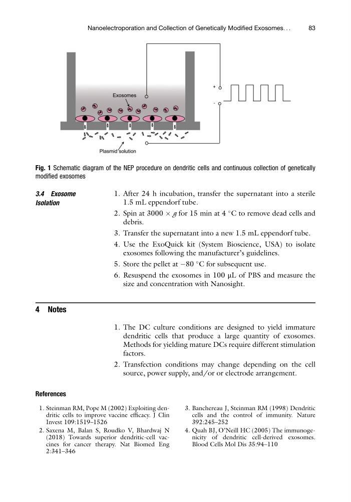
Fig. 1 Schematic diagram of the NEP procedure on dendritic cells and continuous collection of genetically
modified exosomes
Nanoelectroporation and Collection of Genetically Modified Exosomes. . .
83
Isolation
1. After 24 h incubation, transfer the supernatant into a sterile
1.5 mL eppendorf tube.
2. Spin at 3000 Â g for 15 min at 4
C to remove dead cells and
debris.
3. Transfer the supernatant into a new 1.5 mL eppendorf tube.
4. Use the ExoQuick kit (System Bioscience, USA) to isolate
exosomes following the manufacturer’s guidelines.
5. Store the pellet at À80
C for subsequent use.
6. Resuspend the exosomes in 100 μL of PBS and measure the
size and concentration with Nanosight.
4 Notes
1. The DC culture conditions are designed to yield immature
dendritic cells that produce a large quantity of exosomes.
Methods for yielding mature DCs require different stimulation
factors.
2. Transfection conditions may change depending on the cell
source, power supply, and/or or electrode arrangement.
References
1. Steinman RM, Pope M (2002) Exploiting dendritic cells to improve vaccine efficacy. J Clin
Invest 109:1519–1526
2. Saxena M, Balan S, Roudko V, Bhardwaj N
(2018) Towards superior dendritic-cell vaccines for cancer therapy. Nat Biomed Eng
2:341–346
3. Banchereau J, Steinman RM (1998) Dendritic
cells and the control of immunity. Nature
392:245–252
4. Quah BJ, O’Neill HC (2005) The immunogenicity of dendritic cell-derived exosomes.
Blood Cells Mol Dis 35:94–110
Fig. 1 Schematic diagram of the NEP procedure on dendritic cells and continuous collection of genetically
modified exosomes
Nanoelectroporation and Collection of Genetically Modified Exosomes. . .
83