

4. Electroporation are applied between the bottom electrode and
the top rod electrode, which are connected to a power system
(Bio-Rad Gene Pulser).
5. Square-shaped pulses with voltage of 140 V and duration of
10 ms are applied for OSKM factors transfection.
6. Cells are transferred to 6-well plate for culture.
7. YOYO-1 fluorescence intensities are measured on Nikon Ti
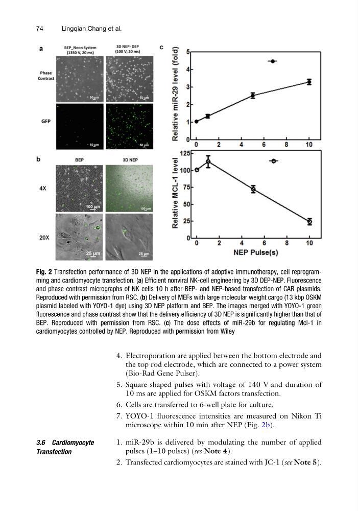
microscope within 10 min after NEP (Fig. 2b).
3.6 Cardiomyocyte
Transfection
1. miR-29b is delivered by modulating the number of applied
pulses (1–10 pulses) (see Note 4).
2. Transfected cardiomyocytes are stained with JC-1 (see Note 5).
Fig. 2 Transfection performance of 3D NEP in the applications of adoptive immunotherapy, cell reprogramming and cardiomyocyte transfection. (a) Efficient nonviral NK-cell engineering by 3D DEP-NEP. Fluorescence
and phase contrast micrographs of NK cells 10 h after BEP- and NEP-based transfection of CAR plasmids.
Reproduced with permission from RSC. (b) Delivery of MEFs with large molecular weight cargo (13 kbp OSKM
plasmid labeled with YOYO-1 dye) using 3D NEP platform and BEP. The images merged with YOYO-1 green
fluorescence and phase contrast show that the delivery efficiency of 3D NEP is significantly higher than that of
BEP. Reproduced with permission from RSC. (c) The dose effects of miR-29b for regulating Mcl-1 in
cardiomyocytes controlled by NEP. Reproduced with permission from Wiley
74
Lingqian Chang et al.
the top rod electrode, which are connected to a power system
(Bio-Rad Gene Pulser).
5. Square-shaped pulses with voltage of 140 V and duration of
10 ms are applied for OSKM factors transfection.
6. Cells are transferred to 6-well plate for culture.
7. YOYO-1 fluorescence intensities are measured on Nikon Ti
microscope within 10 min after NEP (Fig. 2b).
3.6 Cardiomyocyte
Transfection
1. miR-29b is delivered by modulating the number of applied
pulses (1–10 pulses) (see Note 4).
2. Transfected cardiomyocytes are stained with JC-1 (see Note 5).
Fig. 2 Transfection performance of 3D NEP in the applications of adoptive immunotherapy, cell reprogramming and cardiomyocyte transfection. (a) Efficient nonviral NK-cell engineering by 3D DEP-NEP. Fluorescence
and phase contrast micrographs of NK cells 10 h after BEP- and NEP-based transfection of CAR plasmids.
Reproduced with permission from RSC. (b) Delivery of MEFs with large molecular weight cargo (13 kbp OSKM
plasmid labeled with YOYO-1 dye) using 3D NEP platform and BEP. The images merged with YOYO-1 green
fluorescence and phase contrast show that the delivery efficiency of 3D NEP is significantly higher than that of
BEP. Reproduced with permission from RSC. (c) The dose effects of miR-29b for regulating Mcl-1 in
cardiomyocytes controlled by NEP. Reproduced with permission from Wiley
74
Lingqian Chang et al.