

9. Use the Point Click Transfect System to select one cell per
colony and automatically transfect them. A bilevel pulse is
typically applied for the transfection (V 1 ¼ 15 V, t 1 ¼ 0.5 ms,
V 2 ¼ 10 V, t 2 ¼ 2.5 ms, f ¼ 200 Hz, n ¼ 100 pulses). The
amplitude, duration or the number of pulses can be adjusted to
improve the performance (see Note 3).
10. Move to a different region and transfect cells in other colonies
if desired.
11. Disengage the tip and remove the petri dish from the stage.
Replace the media in the petri dish and put it in the incubator.
12. Monitor the cells regularly and add zeocin after 2 days in
culture to select the stably transfected cells.
3.6 Observation,
Colony Isolation
and Cell Line
Generation (Fig. 2c)
1. Observe the cells under the microscope (green channel) to
monitor GFP expression in each colony (see Note 3).
2. Observe the colonies over 2 weeks. Due to zeocin selection
only the stably transfected cells will survive (see Note 4) and
proliferate to form individual colonies at the micropatterned
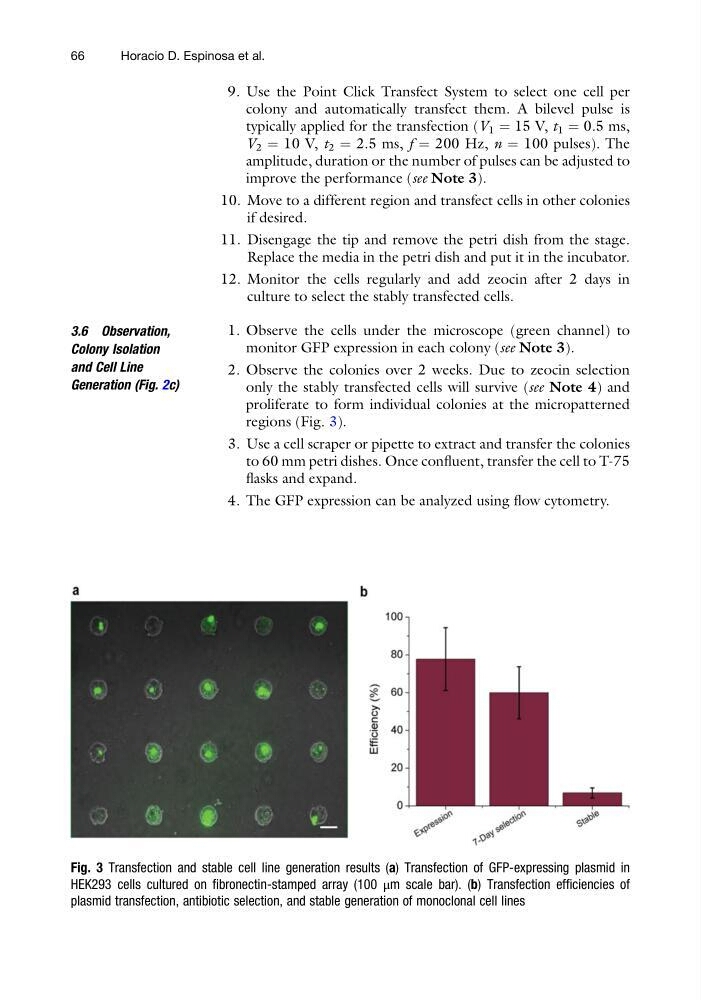
regions (Fig. 3).
3. Use a cell scraper or pipette to extract and transfer the colonies
to 60 mm petri dishes. Once confluent, transfer the cell to T-75
flasks and expand.
4. The GFP expression can be analyzed using flow cytometry.
Fig. 3 Transfection and stable cell line generation results (a) Transfection of GFP-expressing plasmid in
HEK293 cells cultured on fibronectin-stamped array (100 μm scale bar). (b) Transfection efficiencies of
plasmid transfection, antibiotic selection, and stable generation of monoclonal cell lines
66
Horacio D. Espinosa et al.
colony and automatically transfect them. A bilevel pulse is
typically applied for the transfection (V 1 ¼ 15 V, t 1 ¼ 0.5 ms,
V 2 ¼ 10 V, t 2 ¼ 2.5 ms, f ¼ 200 Hz, n ¼ 100 pulses). The
amplitude, duration or the number of pulses can be adjusted to
improve the performance (see Note 3).
10. Move to a different region and transfect cells in other colonies
if desired.
11. Disengage the tip and remove the petri dish from the stage.
Replace the media in the petri dish and put it in the incubator.
12. Monitor the cells regularly and add zeocin after 2 days in
culture to select the stably transfected cells.
3.6 Observation,
Colony Isolation
and Cell Line
Generation (Fig. 2c)
1. Observe the cells under the microscope (green channel) to
monitor GFP expression in each colony (see Note 3).
2. Observe the colonies over 2 weeks. Due to zeocin selection
only the stably transfected cells will survive (see Note 4) and
proliferate to form individual colonies at the micropatterned
regions (Fig. 3).
3. Use a cell scraper or pipette to extract and transfer the colonies
to 60 mm petri dishes. Once confluent, transfer the cell to T-75
flasks and expand.
4. The GFP expression can be analyzed using flow cytometry.
Fig. 3 Transfection and stable cell line generation results (a) Transfection of GFP-expressing plasmid in
HEK293 cells cultured on fibronectin-stamped array (100 μm scale bar). (b) Transfection efficiencies of
plasmid transfection, antibiotic selection, and stable generation of monoclonal cell lines
66
Horacio D. Espinosa et al.