

4. Zeocin—Zeocin antibiotic (InvivoGen, San Diego, CA) at a
final concentration of 400 μg/mL is used in the culture media
for selection of stably transfected cells.
5. Ethanol—70% ethanol is used for cleaning the electrodes.
2.6 Observation
and Cell Line
Generation
1. Fluorescence microscopy—The images are acquired using a
CoolSnap HQ2 camera (Photometrics, Tucson, AZ) mounted
on a Nikon Eclipse Ti microscope. Image acquisition is controlled using NIS Elements software (Nikon, Minato, Japan).
Post processing of images is performed using Fiji image processing package.
2. Colony expansion—Stably transfected colonies are expanded in
60 mm tissue cultures dishes (Corning Inc., Corning, NY) and
T-75 flasks (USA Scientific, Inc., Ocala, FL).
3. Flow cytometry—Characterization of GFP expression and
sorting of GFP expressing cells of interest are done using a
BD FACSAria SORP system at a speed of 1000 cells/s.
3 Methods
3.1 Mold Fabrication
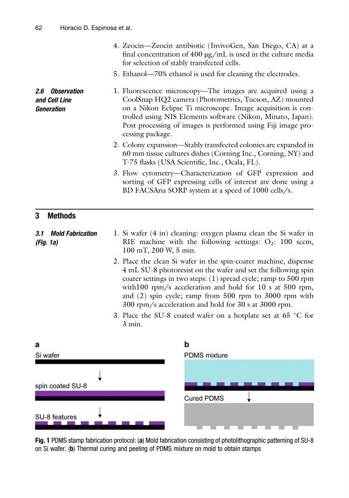
(Fig. 1a)
1. Si wafer (4 in) cleaning: oxygen plasma clean the Si wafer in
RIE machine with the following settings: O 2 : 100 sccm,
100 mT, 200 W, 5 min.
2. Place the clean Si wafer in the spin-coater machine, dispense
4 mL SU-8 photoresist on the wafer and set the following spin
coater settings in two steps: (1) spread cycle; ramp to 500 rpm
with100 rpm/s acceleration and hold for 10 s at 500 rpm,
and (2) spin cycle; ramp from 500 rpm to 3000 rpm with
300 rpm/s acceleration and hold for 30 s at 3000 rpm.
3. Place the SU-8 coated wafer on a hotplate set at 65
C for
3 min.
Fig. 1 PDMS stamp fabrication protocol: (a) Mold fabrication consisting of photolithographic patterning of SU-8
on Si wafer. (b) Thermal curing and peeling of PDMS mixture on mold to obtain stamps
62
Horacio D. Espinosa et al.
final concentration of 400 μg/mL is used in the culture media
for selection of stably transfected cells.
5. Ethanol—70% ethanol is used for cleaning the electrodes.
2.6 Observation
and Cell Line
Generation
1. Fluorescence microscopy—The images are acquired using a
CoolSnap HQ2 camera (Photometrics, Tucson, AZ) mounted
on a Nikon Eclipse Ti microscope. Image acquisition is controlled using NIS Elements software (Nikon, Minato, Japan).
Post processing of images is performed using Fiji image processing package.
2. Colony expansion—Stably transfected colonies are expanded in
60 mm tissue cultures dishes (Corning Inc., Corning, NY) and
T-75 flasks (USA Scientific, Inc., Ocala, FL).
3. Flow cytometry—Characterization of GFP expression and
sorting of GFP expressing cells of interest are done using a
BD FACSAria SORP system at a speed of 1000 cells/s.
3 Methods
3.1 Mold Fabrication
(Fig. 1a)
1. Si wafer (4 in) cleaning: oxygen plasma clean the Si wafer in
RIE machine with the following settings: O 2 : 100 sccm,
100 mT, 200 W, 5 min.
2. Place the clean Si wafer in the spin-coater machine, dispense
4 mL SU-8 photoresist on the wafer and set the following spin
coater settings in two steps: (1) spread cycle; ramp to 500 rpm
with100 rpm/s acceleration and hold for 10 s at 500 rpm,
and (2) spin cycle; ramp from 500 rpm to 3000 rpm with
300 rpm/s acceleration and hold for 30 s at 3000 rpm.
3. Place the SU-8 coated wafer on a hotplate set at 65
C for
3 min.
Fig. 1 PDMS stamp fabrication protocol: (a) Mold fabrication consisting of photolithographic patterning of SU-8
on Si wafer. (b) Thermal curing and peeling of PDMS mixture on mold to obtain stamps
62
Horacio D. Espinosa et al.