

5. As the sealing of the liquid chamber between the two electrodes in the MAE system is done by a PDMS gasket, some
deformation occurred when the top micropillar electrode is
firmly pressed down for closure. Therefore, the actual gap
size between the two electrodes is smaller than the sum of the
measured thickness of the epoxy spacer (10 μm) and the PDMS
gasket (200 μm). In our study, to find out the actual distance
between the two electrodes, we measured the amount of excess
liquid in the connecting channels that is squeezed out of the
liquid chamber when the two plates are closed (see Fig. 3). The
actual gap size is then calculated based on the total volume of
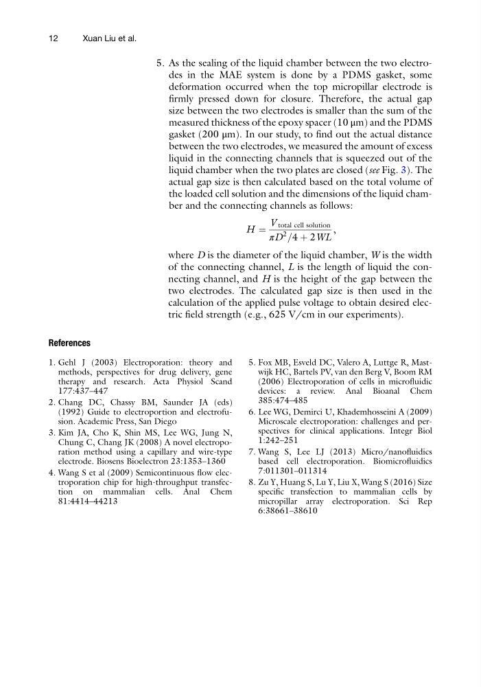
the loaded cell solution and the dimensions of the liquid chamber and the connecting channels as follows:
H ¼
V total cell solution
πD
2
=4 þ 2WL
,
where D is the diameter of the liquid chamber, W is the width
of the connecting channel, L is the length of liquid the connecting channel, and H is the height of the gap between the
two electrodes. The calculated gap size is then used in the
calculation of the applied pulse voltage to obtain desired electric field strength (e.g., 625 V/cm in our experiments).
References
1. Gehl J (2003) Electroporation: theory and
methods, perspectives for drug delivery, gene
therapy and research. Acta Physiol Scand
177:437–447
2. Chang DC, Chassy BM, Saunder JA (eds)
(1992) Guide to electroportion and electrofusion. Academic Press, San Diego
3. Kim JA, Cho K, Shin MS, Lee WG, Jung N,
Chung C, Chang JK (2008) A novel electroporation method using a capillary and wire-type
electrode. Biosens Bioelectron 23:1353–1360
4. Wang S et al (2009) Semicontinuous flow electroporation chip for high-throughput transfection on mammalian cells. Anal Chem
81:4414–44213
5. Fox MB, Esveld DC, Valero A, Luttge R, Mastwijk HC, Bartels PV, van den Berg V, Boom RM
(2006) Electroporation of cells in microfluidic
devices: a review. Anal Bioanal Chem
385:474–485
6. Lee WG, Demirci U, Khademhosseini A (2009)
Microscale electroporation: challenges and perspectives for clinical applications. Integr Biol
1:242–251
7. Wang S, Lee LJ (2013) Micro/nanofluidics
based cell electroporation. Biomicrofluidics
7:011301–011314
8. Zu Y, Huang S, Lu Y, Liu X, Wang S (2016) Size
specific transfection to mammalian cells by
micropillar array electroporation. Sci Rep
6:38661–38610
12
Xuan Liu et al.
deformation occurred when the top micropillar electrode is
firmly pressed down for closure. Therefore, the actual gap
size between the two electrodes is smaller than the sum of the
measured thickness of the epoxy spacer (10 μm) and the PDMS
gasket (200 μm). In our study, to find out the actual distance
between the two electrodes, we measured the amount of excess
liquid in the connecting channels that is squeezed out of the
liquid chamber when the two plates are closed (see Fig. 3). The
actual gap size is then calculated based on the total volume of
the loaded cell solution and the dimensions of the liquid chamber and the connecting channels as follows:
H ¼
V total cell solution
πD
2
=4 þ 2WL
,
where D is the diameter of the liquid chamber, W is the width
of the connecting channel, L is the length of liquid the connecting channel, and H is the height of the gap between the
two electrodes. The calculated gap size is then used in the
calculation of the applied pulse voltage to obtain desired electric field strength (e.g., 625 V/cm in our experiments).
References
1. Gehl J (2003) Electroporation: theory and
methods, perspectives for drug delivery, gene
therapy and research. Acta Physiol Scand
177:437–447
2. Chang DC, Chassy BM, Saunder JA (eds)
(1992) Guide to electroportion and electrofusion. Academic Press, San Diego
3. Kim JA, Cho K, Shin MS, Lee WG, Jung N,
Chung C, Chang JK (2008) A novel electroporation method using a capillary and wire-type
electrode. Biosens Bioelectron 23:1353–1360
4. Wang S et al (2009) Semicontinuous flow electroporation chip for high-throughput transfection on mammalian cells. Anal Chem
81:4414–44213
5. Fox MB, Esveld DC, Valero A, Luttge R, Mastwijk HC, Bartels PV, van den Berg V, Boom RM
(2006) Electroporation of cells in microfluidic
devices: a review. Anal Bioanal Chem
385:474–485
6. Lee WG, Demirci U, Khademhosseini A (2009)
Microscale electroporation: challenges and perspectives for clinical applications. Integr Biol
1:242–251
7. Wang S, Lee LJ (2013) Micro/nanofluidics
based cell electroporation. Biomicrofluidics
7:011301–011314
8. Zu Y, Huang S, Lu Y, Liu X, Wang S (2016) Size
specific transfection to mammalian cells by
micropillar array electroporation. Sci Rep
6:38661–38610
12
Xuan Liu et al.