

transmembrane potentials beyond 700 mV are thought to be
responsible for irreversible damage and lysis of many cellular
structures [7].
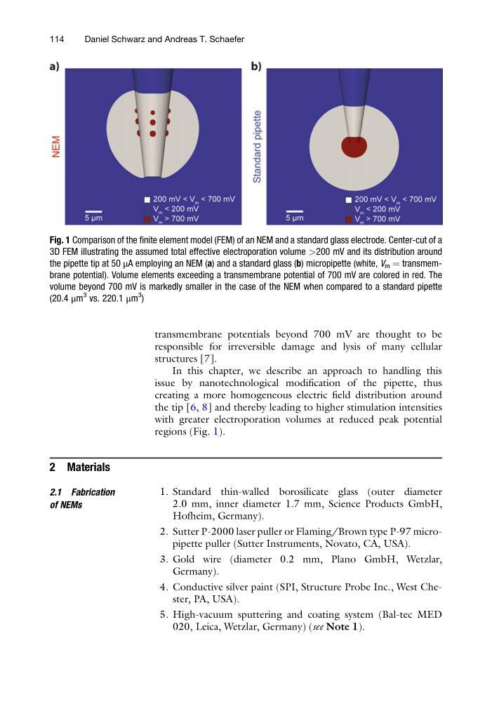
In this chapter, we describe an approach to handling this
issue by nanotechnological modification of the pipette, thus
creating a more homogeneous electric field distribution around
the tip [6, 8] and thereby leading to higher stimulation intensities
with greater electroporation volumes at reduced peak potential
regions (Fig. 1).
2 Materials
2.1 Fabrication
of NEMs
1. Standard thin-walled borosilicate glass (outer diameter
2.0 mm, inner diameter 1.7 mm, Science Products GmbH,
Hofheim, Germany).
2. Sutter P-2000 laser puller or Flaming/Brown type P-97 micropipette puller (Sutter Instruments, Novato, CA, USA).
3. Gold wire (diameter 0.2 mm, Plano GmbH, Wetzlar,
Germany).
4. Conductive silver paint (SPI, Structure Probe Inc., West Chester, PA, USA).
5. High-vacuum sputtering and coating system (Bal-tec MED
020, Leica, Wetzlar, Germany) (see Note 1).
Fig. 1 Comparison of the finite element model (FEM) of an NEM and a standard glass electrode. Center-cut of a
3D FEM illustrating the assumed total effective electroporation volume >200 mV and its distribution around
the pipette tip at 50 μA employing an NEM (a) and a standard glass (b) micropipette (white, V m ¼ transmembrane potential). Volume elements exceeding a transmembrane potential of 700 mV are colored in red. The
volume beyond 700 mV is markedly smaller in the case of the NEM when compared to a standard pipette
(20.4 μm
3 vs. 220.1 μm
3
)
114
Daniel Schwarz and Andreas T. Schaefer
responsible for irreversible damage and lysis of many cellular
structures [7].
In this chapter, we describe an approach to handling this
issue by nanotechnological modification of the pipette, thus
creating a more homogeneous electric field distribution around
the tip [6, 8] and thereby leading to higher stimulation intensities
with greater electroporation volumes at reduced peak potential
regions (Fig. 1).
2 Materials
2.1 Fabrication
of NEMs
1. Standard thin-walled borosilicate glass (outer diameter
2.0 mm, inner diameter 1.7 mm, Science Products GmbH,
Hofheim, Germany).
2. Sutter P-2000 laser puller or Flaming/Brown type P-97 micropipette puller (Sutter Instruments, Novato, CA, USA).
3. Gold wire (diameter 0.2 mm, Plano GmbH, Wetzlar,
Germany).
4. Conductive silver paint (SPI, Structure Probe Inc., West Chester, PA, USA).
5. High-vacuum sputtering and coating system (Bal-tec MED
020, Leica, Wetzlar, Germany) (see Note 1).
Fig. 1 Comparison of the finite element model (FEM) of an NEM and a standard glass electrode. Center-cut of a
3D FEM illustrating the assumed total effective electroporation volume >200 mV and its distribution around
the pipette tip at 50 μA employing an NEM (a) and a standard glass (b) micropipette (white, V m ¼ transmembrane potential). Volume elements exceeding a transmembrane potential of 700 mV are colored in red. The
volume beyond 700 mV is markedly smaller in the case of the NEM when compared to a standard pipette
(20.4 μm
3 vs. 220.1 μm
3
)
114
Daniel Schwarz and Andreas T. Schaefer