

198
M. Dantus and C.L. Kalcic
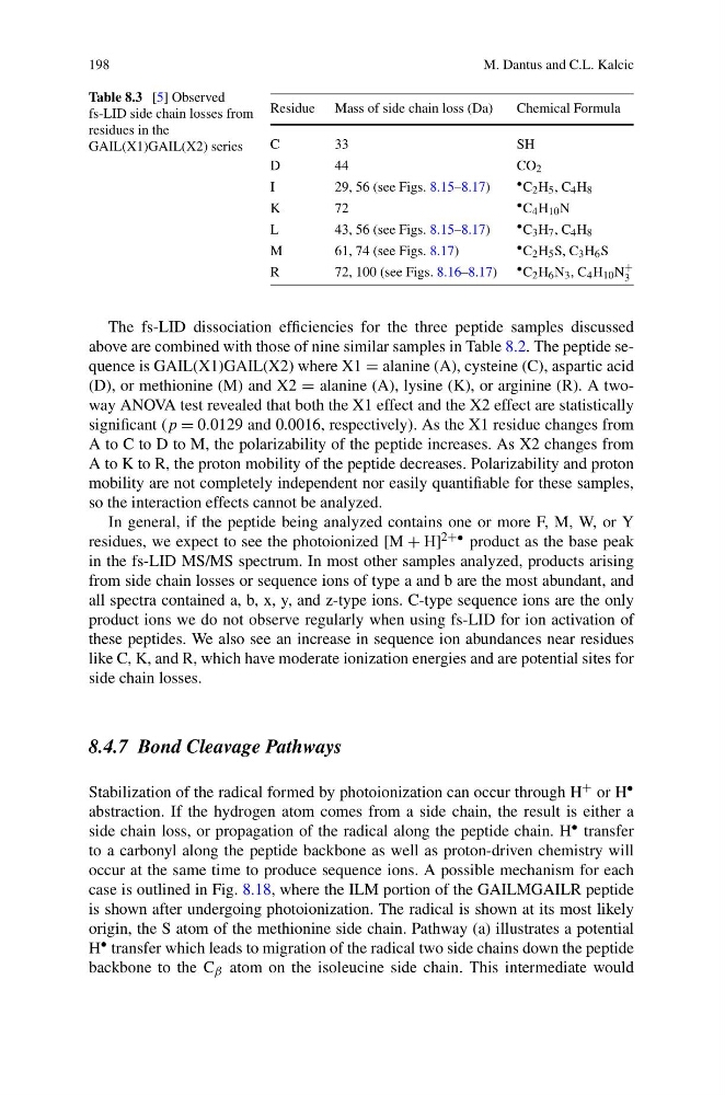
Table 8.3 [5] Observed
fs-LID side chain losses from
residues in the
GAIL(X1)GAIL(X2) series
Residue Mass of side chain loss (Da)
Chemical Formula
C
3 3
S H
D
4 4
C O 2
I
29, 56 (see Figs. 8.15–8.17)
• C 2 H 5 , C 4 H 8
K
7 2
• C 4 H 10 N
L
43, 56 (see Figs. 8.15–8.17)
• C 3 H 7 , C 4 H 8
M
61, 74 (see Figs. 8.17)
• C 2 H 5 S, C 3 H 6 S
R
72, 100 (see Figs. 8.16–8.17) • C 2 H 6 N 3 , C 4 H 10 N
+
3
The fs-LID dissociation efficiencies for the three peptide samples discussed
above are combined with those of nine similar samples in Table 8.2. The peptide sequence is GAIL(X1)GAIL(X2) where X1 = alanine (A), cysteine (C), aspartic acid
(D), or methionine (M) and X2 = alanine (A), lysine (K), or arginine (R). A twoway ANOVA test revealed that both the X1 effect and the X2 effect are statistically
significant (p = 0.0129 and 0.0016, respectively). As the X1 residue changes from
A to C to D to M, the polarizability of the peptide increases. As X2 changes from
A to K to R, the proton mobility of the peptide decreases. Polarizability and proton
mobility are not completely independent nor easily quantifiable for these samples,
so the interaction effects cannot be analyzed.
In general, if the peptide being analyzed contains one or more F, M, W, or Y
residues, we expect to see the photoionized [M + H] 2+• product as the base peak
in the fs-LID MS/MS spectrum. In most other samples analyzed, products arising
from side chain losses or sequence ions of type a and b are the most abundant, and
all spectra contained a, b, x, y, and z-type ions. C-type sequence ions are the only
product ions we do not observe regularly when using fs-LID for ion activation of
these peptides. We also see an increase in sequence ion abundances near residues
like C, K, and R, which have moderate ionization energies and are potential sites for
side chain losses.
8.4.7 Bond Cleavage Pathways
Stabilization of the radical formed by photoionization can occur through H + or H •
abstraction. If the hydrogen atom comes from a side chain, the result is either a
side chain loss, or propagation of the radical along the peptide chain. H • transfer
to a carbonyl along the peptide backbone as well as proton-driven chemistry will
occur at the same time to produce sequence ions. A possible mechanism for each
case is outlined in Fig. 8.18, where the ILM portion of the GAILMGAILR peptide
is shown after undergoing photoionization. The radical is shown at its most likely
origin, the S atom of the methionine side chain. Pathway (a) illustrates a potential
H • transfer which leads to migration of the radical two side chains down the peptide
backbone to the C β atom on the isoleucine side chain. This intermediate would
M. Dantus and C.L. Kalcic
Table 8.3 [5] Observed
fs-LID side chain losses from
residues in the
GAIL(X1)GAIL(X2) series
Residue Mass of side chain loss (Da)
Chemical Formula
C
3 3
S H
D
4 4
C O 2
I
29, 56 (see Figs. 8.15–8.17)
• C 2 H 5 , C 4 H 8
K
7 2
• C 4 H 10 N
L
43, 56 (see Figs. 8.15–8.17)
• C 3 H 7 , C 4 H 8
M
61, 74 (see Figs. 8.17)
• C 2 H 5 S, C 3 H 6 S
R
72, 100 (see Figs. 8.16–8.17) • C 2 H 6 N 3 , C 4 H 10 N
+
3
The fs-LID dissociation efficiencies for the three peptide samples discussed
above are combined with those of nine similar samples in Table 8.2. The peptide sequence is GAIL(X1)GAIL(X2) where X1 = alanine (A), cysteine (C), aspartic acid
(D), or methionine (M) and X2 = alanine (A), lysine (K), or arginine (R). A twoway ANOVA test revealed that both the X1 effect and the X2 effect are statistically
significant (p = 0.0129 and 0.0016, respectively). As the X1 residue changes from
A to C to D to M, the polarizability of the peptide increases. As X2 changes from
A to K to R, the proton mobility of the peptide decreases. Polarizability and proton
mobility are not completely independent nor easily quantifiable for these samples,
so the interaction effects cannot be analyzed.
In general, if the peptide being analyzed contains one or more F, M, W, or Y
residues, we expect to see the photoionized [M + H] 2+• product as the base peak
in the fs-LID MS/MS spectrum. In most other samples analyzed, products arising
from side chain losses or sequence ions of type a and b are the most abundant, and
all spectra contained a, b, x, y, and z-type ions. C-type sequence ions are the only
product ions we do not observe regularly when using fs-LID for ion activation of
these peptides. We also see an increase in sequence ion abundances near residues
like C, K, and R, which have moderate ionization energies and are potential sites for
side chain losses.
8.4.7 Bond Cleavage Pathways
Stabilization of the radical formed by photoionization can occur through H + or H •
abstraction. If the hydrogen atom comes from a side chain, the result is either a
side chain loss, or propagation of the radical along the peptide chain. H • transfer
to a carbonyl along the peptide backbone as well as proton-driven chemistry will
occur at the same time to produce sequence ions. A possible mechanism for each
case is outlined in Fig. 8.18, where the ILM portion of the GAILMGAILR peptide
is shown after undergoing photoionization. The radical is shown at its most likely
origin, the S atom of the methionine side chain. Pathway (a) illustrates a potential
H • transfer which leads to migration of the radical two side chains down the peptide
backbone to the C β atom on the isoleucine side chain. This intermediate would