

196
M. Dantus and C.L. Kalcic
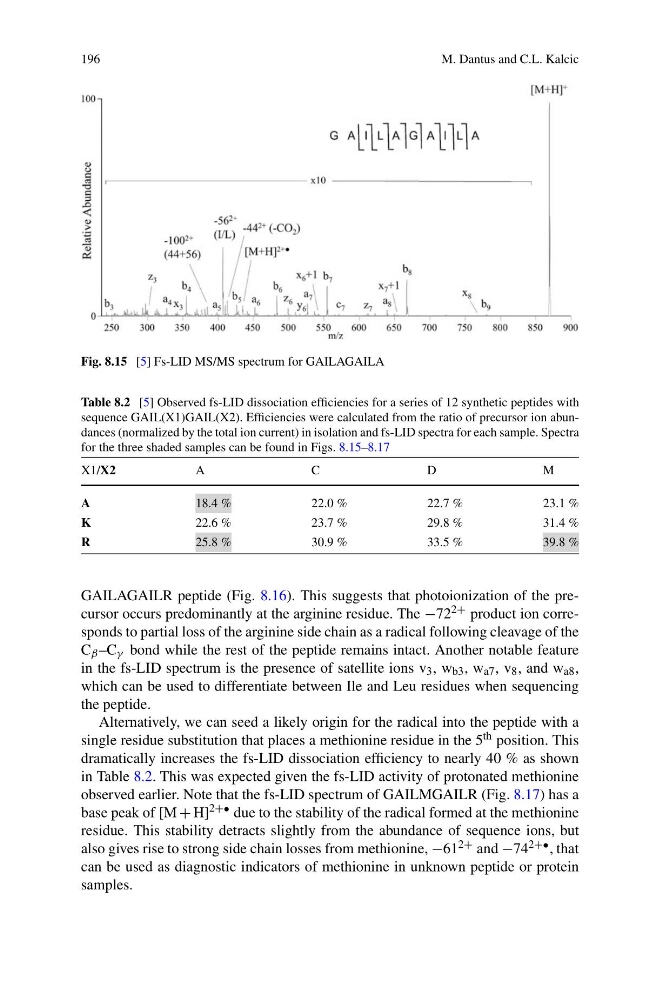
Fig. 8.15 [5] Fs-LID MS/MS spectrum for GAILAGAILA
Table 8.2 [5] Observed fs-LID dissociation efficiencies for a series of 12 synthetic peptides with
sequence GAIL(X1)GAIL(X2). Efficiencies were calculated from the ratio of precursor ion abundances (normalized by the total ion current) in isolation and fs-LID spectra for each sample. Spectra
for the three shaded samples can be found in Figs. 8.15–8.17
X1/X2
A
C
D
M
A
18.4 %
22.0 %
22.7 %
23.1 %
K
22.6 %
23.7 %
29.8 %
31.4 %
R
25.8 %
30.9 %
33.5 %
39.8 %
GAILAGAILR peptide (Fig. 8.16). This suggests that photoionization of the precursor occurs predominantly at the arginine residue. The −72 2+ product ion corresponds to partial loss of the arginine side chain as a radical following cleavage of the
C β –C γ bond while the rest of the peptide remains intact. Another notable feature
in the fs-LID spectrum is the presence of satellite ions v 3 , w b3 , w a7 , v 8 , and w a8 ,
which can be used to differentiate between Ile and Leu residues when sequencing
the peptide.
Alternatively, we can seed a likely origin for the radical into the peptide with a
single residue substitution that places a methionine residue in the 5 th position. This
dramatically increases the fs-LID dissociation efficiency to nearly 40 % as shown
in Table 8.2. This was expected given the fs-LID activity of protonated methionine
observed earlier. Note that the fs-LID spectrum of GAILMGAILR (Fig. 8.17) has a
base peak of [M + H] 2+• due to the stability of the radical formed at the methionine
residue. This stability detracts slightly from the abundance of sequence ions, but
also gives rise to strong side chain losses from methionine, −61 2+ and −74 2+• , that
can be used as diagnostic indicators of methionine in unknown peptide or protein
samples.
M. Dantus and C.L. Kalcic
Fig. 8.15 [5] Fs-LID MS/MS spectrum for GAILAGAILA
Table 8.2 [5] Observed fs-LID dissociation efficiencies for a series of 12 synthetic peptides with
sequence GAIL(X1)GAIL(X2). Efficiencies were calculated from the ratio of precursor ion abundances (normalized by the total ion current) in isolation and fs-LID spectra for each sample. Spectra
for the three shaded samples can be found in Figs. 8.15–8.17
X1/X2
A
C
D
M
A
18.4 %
22.0 %
22.7 %
23.1 %
K
22.6 %
23.7 %
29.8 %
31.4 %
R
25.8 %
30.9 %
33.5 %
39.8 %
GAILAGAILR peptide (Fig. 8.16). This suggests that photoionization of the precursor occurs predominantly at the arginine residue. The −72 2+ product ion corresponds to partial loss of the arginine side chain as a radical following cleavage of the
C β –C γ bond while the rest of the peptide remains intact. Another notable feature
in the fs-LID spectrum is the presence of satellite ions v 3 , w b3 , w a7 , v 8 , and w a8 ,
which can be used to differentiate between Ile and Leu residues when sequencing
the peptide.
Alternatively, we can seed a likely origin for the radical into the peptide with a
single residue substitution that places a methionine residue in the 5 th position. This
dramatically increases the fs-LID dissociation efficiency to nearly 40 % as shown
in Table 8.2. This was expected given the fs-LID activity of protonated methionine
observed earlier. Note that the fs-LID spectrum of GAILMGAILR (Fig. 8.17) has a
base peak of [M + H] 2+• due to the stability of the radical formed at the methionine
residue. This stability detracts slightly from the abundance of sequence ions, but
also gives rise to strong side chain losses from methionine, −61 2+ and −74 2+• , that
can be used as diagnostic indicators of methionine in unknown peptide or protein
samples.