

11 Segmentation and Visualization of Drusen …
311
that the method may be effective for the GA segmentation in SD-OCT images. This
segmentation algorithm can also be used to extract and assess GA quantitative features in longitudinal OCT studies, such as the area and extent of GA. A performance
comparison of our algorithm with a commercially-available GA segmentation software program suggests that our algorithm provides more accurate GA segmentations
than the commercial software.
11.3.2 Automated Geographic Atrophy Segmentation
for SD-OCT Images Using Region-Based C-V Model
via Local Similarity Factor
11.3.2.1 Methods
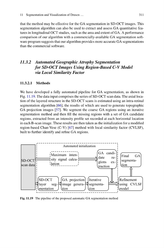
We have developed a fully automated pipeline for GA segmentation, as shown in
Fig. 11.19. The data input comprises the series of SD-OCT scan data. The axial location of the layered structure in the SD-OCT scans is estimated using an intra-retinal
segmentation algorithm [66], the results of which are used to generate topographic
GA projection images [57]. We segment the coarse GA regions using an iterative
segmentation method and then fill the missing regions with a set of GA candidate
regions, extracted from an intensity profile set recorded at each horizontal location
in each B-scan image. These results are then taken as the initialization for a modified
region-based Chan-Vese (C-V) [67] method with local similarity factor (CVLSF),
built to further identify and refine GA regions.
Automated initialization
SD-OCT
scan data
SD-OCT
layer segmentation
GA projection
image generation
Iterative
segmentation
Refinement
using CVLSF
model
Final
GA
segmentation
Maximum intensity signal calculation
GA candidate
regions extraction
Fig. 11.19 The pipeline of the proposed automatic GA segmentation method
311
that the method may be effective for the GA segmentation in SD-OCT images. This
segmentation algorithm can also be used to extract and assess GA quantitative features in longitudinal OCT studies, such as the area and extent of GA. A performance
comparison of our algorithm with a commercially-available GA segmentation software program suggests that our algorithm provides more accurate GA segmentations
than the commercial software.
11.3.2 Automated Geographic Atrophy Segmentation
for SD-OCT Images Using Region-Based C-V Model
via Local Similarity Factor
11.3.2.1 Methods
We have developed a fully automated pipeline for GA segmentation, as shown in
Fig. 11.19. The data input comprises the series of SD-OCT scan data. The axial location of the layered structure in the SD-OCT scans is estimated using an intra-retinal
segmentation algorithm [66], the results of which are used to generate topographic
GA projection images [57]. We segment the coarse GA regions using an iterative
segmentation method and then fill the missing regions with a set of GA candidate
regions, extracted from an intensity profile set recorded at each horizontal location
in each B-scan image. These results are then taken as the initialization for a modified
region-based Chan-Vese (C-V) [67] method with local similarity factor (CVLSF),
built to further identify and refine GA regions.
Automated initialization
SD-OCT
scan data
SD-OCT
layer segmentation
GA projection
image generation
Iterative
segmentation
Refinement
using CVLSF
model
Final
GA
segmentation
Maximum intensity signal calculation
GA candidate
regions extraction
Fig. 11.19 The pipeline of the proposed automatic GA segmentation method