

302
Q. Chen et al.
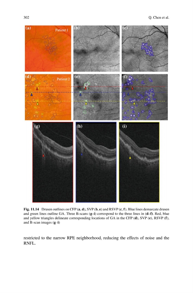
Fig. 11.14 Drusen outlines on CFP (a, d), SVP (b, e) and RSVP (c, f). Blue lines demarcate drusen
and green lines outline GA. Three B-scans (g–i) correspond to the three lines in (d–f). Red, blue
and yellow triangles delineate corresponding locations of GA in the CFP (d), SVP (e), RSVP (f),
and B-scan images (g–i)
restricted to the narrow RPE neighborhood, reducing the effects of noise and the
RNFL.
Q. Chen et al.
Fig. 11.14 Drusen outlines on CFP (a, d), SVP (b, e) and RSVP (c, f). Blue lines demarcate drusen
and green lines outline GA. Three B-scans (g–i) correspond to the three lines in (d–f). Red, blue
and yellow triangles delineate corresponding locations of GA in the CFP (d), SVP (e), RSVP (f),
and B-scan images (g–i)
restricted to the narrow RPE neighborhood, reducing the effects of noise and the
RNFL.