

296
Q. Chen et al.
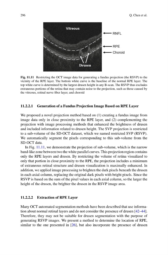
Fig. 11.11 Restricting the OCT image data for generating a fundus projection (the RSVP) to the
vicinity of the RPE layer. The bottom white curve is the baseline of the normal RPE layer. The
top white curve is determined by the largest drusen height in any B-scan. The RSVP thus excludes
extraneous portions of the retina that may contain noise to the projection, such as those caused by
the vitreous, retinal nerve fiber layer, and choroid
11.2.2.1 Generation of a Fundus Projection Image Based on RPE Layer
We proposed a novel projection method based on (1) creating a fundus image from
image data only in close proximity to the RPE layer, and (2) complementing the
projection with image processing methods that enhanced the brightness of drusen
and included information related to drusen height. The SVP projection is restricted
to a sub-volume of the SD-OCT dataset, which we named restricted SVP (RSVP).
We automatically segment the pixels corresponding to this sub-volume from the
SD-OCT data.
In Fig. 11.11, we demonstrate the projection of sub-volume, which is the narrow
band-like zone between two the white parallel curves. This projection region contains
only the RPE layers and drusen. By restricting the volume of retina visualized to
only that portion in close proximity to the RPE, the projection includes a minimum
of extraneous retinal structure and drusen visualization is maximally enhanced. In
addition, we applied image processing to brighten the dark pixels beneath the drusen
in each axial column, replacing the original dark pixels with bright pixels. Since the
RSVP is based on the sum of the pixel values in each axial column, so the larger the
height of the drusen, the brighter the drusen in the RSVP image area.
11.2.2.2 Extraction of RPE Layer
Many OCT automated segmentation methods have been described that use information about normal retinal layers and do not consider the presence of drusen [42–44].
Therefore, they may not be suitable for drusen segmentation with the purpose of
generating RSVP images. We present a method to determine the location of RPE,
similar to the one presented in [26], but also incorporate the presence of drusen
Q. Chen et al.
Fig. 11.11 Restricting the OCT image data for generating a fundus projection (the RSVP) to the
vicinity of the RPE layer. The bottom white curve is the baseline of the normal RPE layer. The
top white curve is determined by the largest drusen height in any B-scan. The RSVP thus excludes
extraneous portions of the retina that may contain noise to the projection, such as those caused by
the vitreous, retinal nerve fiber layer, and choroid
11.2.2.1 Generation of a Fundus Projection Image Based on RPE Layer
We proposed a novel projection method based on (1) creating a fundus image from
image data only in close proximity to the RPE layer, and (2) complementing the
projection with image processing methods that enhanced the brightness of drusen
and included information related to drusen height. The SVP projection is restricted
to a sub-volume of the SD-OCT dataset, which we named restricted SVP (RSVP).
We automatically segment the pixels corresponding to this sub-volume from the
SD-OCT data.
In Fig. 11.11, we demonstrate the projection of sub-volume, which is the narrow
band-like zone between two the white parallel curves. This projection region contains
only the RPE layers and drusen. By restricting the volume of retina visualized to
only that portion in close proximity to the RPE, the projection includes a minimum
of extraneous retinal structure and drusen visualization is maximally enhanced. In
addition, we applied image processing to brighten the dark pixels beneath the drusen
in each axial column, replacing the original dark pixels with bright pixels. Since the
RSVP is based on the sum of the pixel values in each axial column, so the larger the
height of the drusen, the brighter the drusen in the RSVP image area.
11.2.2.2 Extraction of RPE Layer
Many OCT automated segmentation methods have been described that use information about normal retinal layers and do not consider the presence of drusen [42–44].
Therefore, they may not be suitable for drusen segmentation with the purpose of
generating RSVP images. We present a method to determine the location of RPE,
similar to the one presented in [26], but also incorporate the presence of drusen