

10 Layer Segmentation and Analysis for Retina with Diseases
255
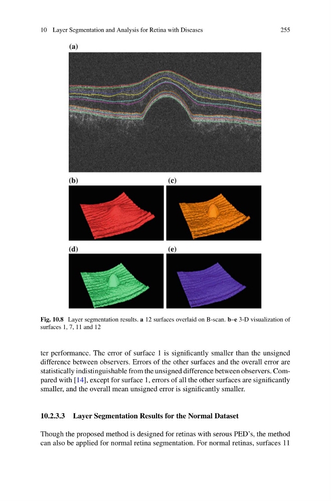
Fig. 10.8 Layer segmentation results. a 12 surfaces overlaid on B-scan. b–e 3-D visualization of
surfaces 1, 7, 11 and 12
ter performance. The error of surface 1 is significantly smaller than the unsigned
difference between observers. Errors of the other surfaces and the overall error are
statistically indistinguishable from the unsigned difference between observers. Compared with [14], except for surface 1, errors of all the other surfaces are significantly
smaller, and the overall mean unsigned error is significantly smaller.
10.2.3.3 Layer Segmentation Results for the Normal Dataset
Though the proposed method is designed for retinas with serous PED’s, the method
can also be applied for normal retina segmentation. For normal retinas, surfaces 11
255
Fig. 10.8 Layer segmentation results. a 12 surfaces overlaid on B-scan. b–e 3-D visualization of
surfaces 1, 7, 11 and 12
ter performance. The error of surface 1 is significantly smaller than the unsigned
difference between observers. Errors of the other surfaces and the overall error are
statistically indistinguishable from the unsigned difference between observers. Compared with [14], except for surface 1, errors of all the other surfaces are significantly
smaller, and the overall mean unsigned error is significantly smaller.
10.2.3.3 Layer Segmentation Results for the Normal Dataset
Though the proposed method is designed for retinas with serous PED’s, the method
can also be applied for normal retina segmentation. For normal retinas, surfaces 11