

216
K. K. Vupparaboina et al.
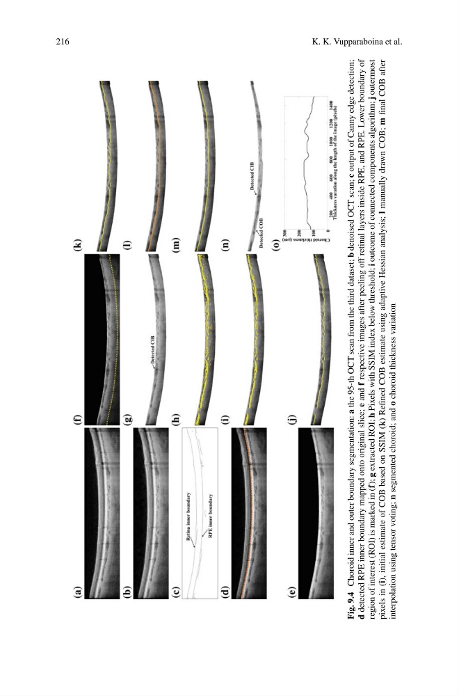
Fig. 9.4
Choroid inner and outer boundary segmentation: a
the 95-th OCT scan from the third dataset; b
denoised OCT scan; c
output of Canny edge detection;
d
detected RPE inner boundary mapped onto original slice; e
and f
respective images after peeling off retinal layers inside RPE, and RPE. Lower boundary of
region of interest (ROI) is marked in (f); g
extracted ROI; h
Pixels with SSIM index below threshold; i
outcome of connected components algorithm; j
outermost
pixels in (i), initial estimate of COB based on SSIM (k) Refined COB estimate using adaptive Hessian analysis; l
manually drawn COB; m
final COB after
interpolation using tensor voting; n
segmented choroid; and o
choroid thickness variation
K. K. Vupparaboina et al.
Fig. 9.4
Choroid inner and outer boundary segmentation: a
the 95-th OCT scan from the third dataset; b
denoised OCT scan; c
output of Canny edge detection;
d
detected RPE inner boundary mapped onto original slice; e
and f
respective images after peeling off retinal layers inside RPE, and RPE. Lower boundary of
region of interest (ROI) is marked in (f); g
extracted ROI; h
Pixels with SSIM index below threshold; i
outcome of connected components algorithm; j
outermost
pixels in (i), initial estimate of COB based on SSIM (k) Refined COB estimate using adaptive Hessian analysis; l
manually drawn COB; m
final COB after
interpolation using tensor voting; n
segmented choroid; and o
choroid thickness variation