

8 Segmentation of Optic Disc and Cup-to-Disc Ratio Quantification …
207
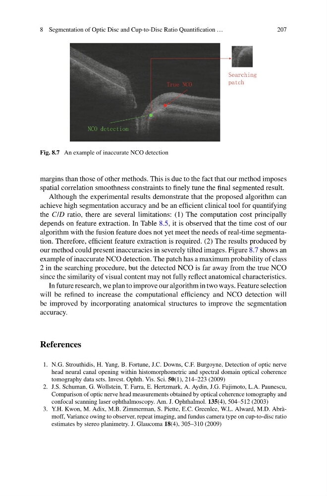
Fig. 8.7 An example of inaccurate NCO detection
margins than those of other methods. This is due to the fact that our method imposes
spatial correlation smoothness constraints to finely tune the final segmented result.
Although the experimental results demonstrate that the proposed algorithm can
achieve high segmentation accuracy and be an efficient clinical tool for quantifying
the C/D ratio, there are several limitations: (1) The computation cost principally
depends on feature extraction. In Table 8.5, it is observed that the time cost of our
algorithm with the fusion feature does not yet meet the needs of real-time segmentation. Therefore, efficient feature extraction is required. (2) The results produced by
our method could present inaccuracies in severely tilted images. Figure 8.7 shows an
example of inaccurate NCO detection. The patch has a maximum probability of class
2 in the searching procedure, but the detected NCO is far away from the true NCO
since the similarity of visual content may not fully reflect anatomical characteristics.
In future research, we plan to improve our algorithm in two ways. Feature selection
will be refined to increase the computational efficiency and NCO detection will
be improved by incorporating anatomical structures to improve the segmentation
accuracy.
References
1. N.G. Strouthidis, H. Yang, B. Fortune, J.C. Downs, C.F. Burgoyne, Detection of optic nerve
head neural canal opening within histomorphometric and spectral domain optical coherence
tomography data sets. Invest. Ophth. Vis. Sci. 50(1), 214–223 (2009)
2. J.S. Schuman, G. Wollstein, T. Farra, E. Hertzmark, A. Aydin, J.G. Fujimoto, L.A. Paunescu,
Comparison of optic nerve head measurements obtained by optical coherence tomography and
confocal scanning laser ophthalmoscopy. Am. J. Ophthalmol. 135(4), 504–512 (2003)
3. Y.H. Kwon, M. Adix, M.B. Zimmerman, S. Piette, E.C. Greenlee, W.L. Alward, M.D. Abràmoff, Variance owing to observer, repeat imaging, and fundus camera type on cup-to-disc ratio
estimates by stereo planimetry. J. Glaucoma 18(4), 305–310 (2009)
207
Fig. 8.7 An example of inaccurate NCO detection
margins than those of other methods. This is due to the fact that our method imposes
spatial correlation smoothness constraints to finely tune the final segmented result.
Although the experimental results demonstrate that the proposed algorithm can
achieve high segmentation accuracy and be an efficient clinical tool for quantifying
the C/D ratio, there are several limitations: (1) The computation cost principally
depends on feature extraction. In Table 8.5, it is observed that the time cost of our
algorithm with the fusion feature does not yet meet the needs of real-time segmentation. Therefore, efficient feature extraction is required. (2) The results produced by
our method could present inaccuracies in severely tilted images. Figure 8.7 shows an
example of inaccurate NCO detection. The patch has a maximum probability of class
2 in the searching procedure, but the detected NCO is far away from the true NCO
since the similarity of visual content may not fully reflect anatomical characteristics.
In future research, we plan to improve our algorithm in two ways. Feature selection
will be refined to increase the computational efficiency and NCO detection will
be improved by incorporating anatomical structures to improve the segmentation
accuracy.
References
1. N.G. Strouthidis, H. Yang, B. Fortune, J.C. Downs, C.F. Burgoyne, Detection of optic nerve
head neural canal opening within histomorphometric and spectral domain optical coherence
tomography data sets. Invest. Ophth. Vis. Sci. 50(1), 214–223 (2009)
2. J.S. Schuman, G. Wollstein, T. Farra, E. Hertzmark, A. Aydin, J.G. Fujimoto, L.A. Paunescu,
Comparison of optic nerve head measurements obtained by optical coherence tomography and
confocal scanning laser ophthalmoscopy. Am. J. Ophthalmol. 135(4), 504–512 (2003)
3. Y.H. Kwon, M. Adix, M.B. Zimmerman, S. Piette, E.C. Greenlee, W.L. Alward, M.D. Abràmoff, Variance owing to observer, repeat imaging, and fundus camera type on cup-to-disc ratio
estimates by stereo planimetry. J. Glaucoma 18(4), 305–310 (2009)