

182
E. Gao et al.
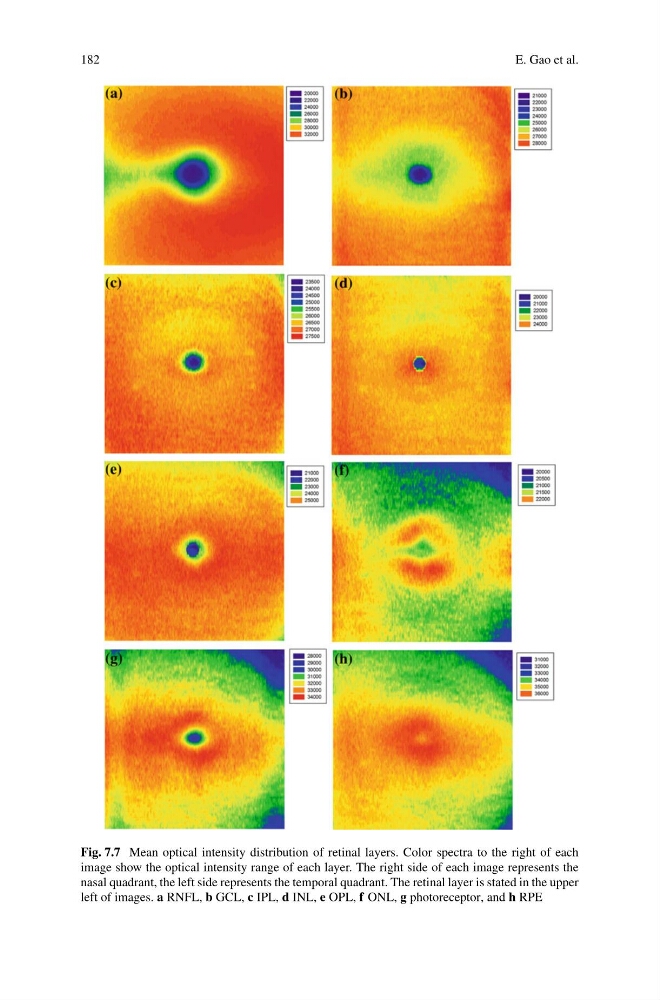
Fig. 7.7 Mean optical intensity distribution of retinal layers. Color spectra to the right of each
image show the optical intensity range of each layer. The right side of each image represents the
nasal quadrant, the left side represents the temporal quadrant. The retinal layer is stated in the upper
left of images. a RNFL, b GCL, c IPL, d INL, e OPL, f ONL, g photoreceptor, and h RPE
E. Gao et al.
Fig. 7.7 Mean optical intensity distribution of retinal layers. Color spectra to the right of each
image show the optical intensity range of each layer. The right side of each image represents the
nasal quadrant, the left side represents the temporal quadrant. The retinal layer is stated in the upper
left of images. a RNFL, b GCL, c IPL, d INL, e OPL, f ONL, g photoreceptor, and h RPE